A mouth ulcer, sore, or unusual white or red patch may seem harmless, so often people wait for it to heal on its own. However, changes that last longer than two weeks should not be ignored1. Mouth cancer is an important health concern in India. About 1,41,342 people are diagnosed with oral cancer, with around 10 cases reported per 100,000 people. Tobacco use is one of the major preventable factors linked to oral cancer; about 80% of these cases could potentially be avoided by not using or quitting tobacco2. This article explains the early signs of mouth cancer and when to seek medical advice.
Mouth cancer, also called oral cancer, is cancer that starts in the tissues of the mouth. It may affect the lips, tongue, gums, inner lining of the cheeks and lips, floor of the mouth, or roof of the mouth. If it is not found and treated early, it can grow into nearby tissues and may spread to other parts of the body1.
The following are some facts about mouth cancer:
Mouth cancers can originate from different types of cells or tissues. Common types include:
Mouth cancer can cause several symptoms. Possible early symptoms of mouth cancer include1:
Mouth cancer develops when cells in the mouth change in a way that makes them grow and divide out of control. Factors that can increase the risk include:
A risk factor is something that can make a person more likely to develop a disease. The following are some risk factors for mouth cancer:
Note: These factors can increase the risk of mouth cancer, but having one or more of them does not mean that a person will definitely develop the disease. If you have risk factors and notice possible mouth cancer symptoms, speak with a dentist or doctor.
Also Read: Is Cancer a Genetic Disease? Hereditary Risk & Genetic Testing Guide
Finding and treating mouth cancer at an early stage is important for several reasons:
Doctors use a combination of examination and tests to diagnose mouth cancer. The process may include:
Note: Depending on the suspected cancer and its stage, doctors may recommend other tests to determine how far the disease has spread and to help plan treatment.
Treatment for oral cancer depends on the type, location, and stage of the cancer, as well as the person’s general health. Common mouth cancer treatment options include:
Along with medical treatment, some daily habits may help people cope with mouth cancer and treatment side effects.
Note: These tips are for general support and should not replace medical advice. Your doctor or dentist may recommend different food choices, oral care, or daily activities depending on your treatment and individual needs.
You may be able to lower the risk of oral cancer by following these steps:
Note: These tips are meant to help you stay informed and lower your risk of mouth cancer. However, if you feel any suspicious changes in your mouth, it is important to consult a doctor, even if you have been following these tips.
Also Read: Tongue Cancer: Symptoms, Causes, Types, Stages and Treatment Options
See a doctor or dentist if a sore or patch in the mouth does not heal within two weeks, or if you notice an unusual lump or wart, unexplained bleeding, pain, numbness, a loose tooth, dentures that suddenly fit poorly, trouble chewing or swallowing, difficulty moving the tongue or jaw, a lump in the neck, or unexplained weight loss along with mouth changes1,3. These symptoms can happen for reasons other than cancer, but a check-up is the safest way to find the cause.
Also Read: Types of Cancer: Symptoms, Early Signs, Diagnosis & More
Knowing the early-stage symptoms of mouth cancer can help you notice unusual changes sooner. Avoiding tobacco and areca nut, limiting alcohol, protecting the lips from sun exposure, and maintaining good oral health may help lower the risk. If any mouth change lasts for more than two weeks, do not ignore it. Speak with a doctor or dentist. Early medical attention can support timely diagnosis and treatment.
Mouth cancer may be noticed early by paying attention to changes such as sores that do not heal, unusual red or white patches, lumps on the neck, bleeding, or persistent pain. If any of these changes last for a few weeks, it is important to consult a dentist/doctor1,3.
In the beginning, mouth cancer may not always be painful. Some people notice a sore that does not heal, a red or white patch, a lump, or a thickened area in the mouth1.
Yes, mouth cancer can often be treated successfully, especially when it is found early. The chances of successful treatment depend on factors such as the cancer’s stage, location, spread, type, and paient’s response to treatment and overall health12.
Mouth cancer may not cause pain in the beginning, and some people may notice only a sore, patch, lump or other unusual change. Persistent pain or discomfort can develop as the condition progresses12.
Yes, chewing tobacco exposes the mouth to harmful substances that can damage oral tissues and increase the risk of developing mouth cancer6,12.
Yes, mouth cancer can spread to nearby tissues and lymph nodes and, in advanced cases, may reach other parts of the body. Early diagnosis and treatment can help reduce the risk of further spread.
1. Oral cancer. MedlinePlus. 2025. Accessed August 12, 2026. https://medlineplus.gov/ency/article/001035.htm
2. New genome-wide study from Tata Memorial Centre reveals: “Why do some tobacco chewers develop oral cancer early? Government of India. 2025. Accessed August 12, 2026. https://www.pib.gov.in/PressReleaseDetail.aspx?PRID=2196291®=1&lang=1
3. Oral cancer: Mouth Cancer. MedlinePlus. 2024. Accessed August 12, 2026. https://medlineplus.gov/oralcancer.html
4. Types of mouth and oropharyngeal cancers. Cancer Research UK. 2024. Accessed August 12, 2026. https://www.cancerresearchuk.org/about-cancer/mouth-cancer/stages-types-grades/types-grades
5. Zito PM, Brizuela M, Mazzoni T. Oral melanoma. In: StatPearls. StatPearls Publishing; 2023. Accessed August 12, 2026. http://www.ncbi.nlm.nih.gov/books/NBK513276/
6. Oral Cancer. NIDCR. 2024. Accessed August 12, 2026. https://www.nidcr.nih.gov/health-info/oral-cancer
7. Warnakulasuriya S, Trivedy C, Peters TJ. Areca nut use: an independent risk factor for oral cancer. BMJ. 2002;324(7341):799-800. doi:10.1136/bmj.324.7341.799 https://pubmed.ncbi.nlm.nih.gov/11934759/
8. Feng L, Wang L. Effects of alcohol on the morphological and structural changes in oral mucosa – PMC. 2013. doi:10.12669/pjms.294.3696 https://pmc.ncbi.nlm.nih.gov/articles/PMC3817782/
9. HPV and oropharyngeal cancer | cancer. CDC. 2024. Accessed August 12, 2026. https://www.cdc.gov/cancer/hpv/oropharyngeal-cancer.html
10. Oral cavity and oropharyngeal cancer causes, risk factors, and prevention. American Cancer Society. 2026. Accessed August 12, 2026. https://www.cancer.org/cancer/types/oral-cavity-and-oropharyngeal-cancer/causes-risks-prevention.html
11. Mathur R, Singhavi HR, Malik A, Nair S, Chaturvedi P. Role of poor oral hygiene in causation of oral cancer—a review of literature. Indian J Surg Oncol. 2018;10(1):184-195. doi:10.1007/s13193-018-0836-5 https://pubmed.ncbi.nlm.nih.gov/30948897/
12. Sankaranarayanan R, Ramadas K, Amarasinghe H, Subramanian S, Johnson N. Oral cancer: Prevention, early detection, and treatment. In: Gelband H, Jha P, Sankaranarayanan R, Horton S, eds. Cancer: Disease Control Priorities, Third Edition (Volume 3). The International Bank for Reconstruction and Development / The World Bank; 2015. Accessed August 12, 2026. http://www.ncbi.nlm.nih.gov/books/NBK343649/
13. Singh AG, Chaukar D, Gupta S, et al. A prospective study to determine the cost of illness for oral cancer in India. Ecancermedicalscience. 2021;15:1252. doi:10.3332/ecancer.2021.1252 https://pubmed.ncbi.nlm.nih.gov/34267808/
14. Macey R, Walsh T, Brocklehurst P, et al. Diagnostic tests for oral cancer and potentially malignant disorders in patients presenting with clinically evident lesions. Cochrane Database Syst Rev. 2015;2015(5):CD010276. doi:10.1002/14651858.CD010276.pub2 https://pubmed.ncbi.nlm.nih.gov/34282854/
15. Early Detection, Diagnosis and Staging – Oral Cancer Foundation | Information and Resources about Oral Head and Neck Cancer. Accessed August 12, 2026. https://oralcancerfoundation.org/cdc/early-detection-diagnosis-staging/
16. Treatment for mouth cancer – NHS. 2023. Accessed August 12, 2026. https://www.nhs.uk/conditions/mouth-cancer/treatment/
17. Oral complications of cancer therapies. National Cancer Institute. 2024. Accessed August 12, 2026. https://www.cancer.gov/about-cancer/treatment/side-effects/mouth-throat/oral-complications-pdq
18. Distant metastasis from oral cancer: A review and molecular biologic aspect. PMC. 2016. Accessed August 12, 2026. https://pmc.ncbi.nlm.nih.gov/articles/PMC4981925/
19. Asmin PK, Nusrath F, Divakar DD. Occurrence and distribution of cancers with emphasis upon oral cancers in registered oncology institutes of South India–A retrospective study. Indian Journal of Community Medicine: Official Publication of Indian Association of Preventive & Social Medicine. 2024 Jan 12;49(1):120. https://pubmed.ncbi.nlm.nih.gov/38425965/
Disclaimer: The information provided here is for educational/awareness purposes only and is not intended to be a substitute for medical treatment by a healthcare professional and should not be relied upon to diagnose or treat any medical condition. The reader should consult a registered medical practitioner to determine the appropriateness of the information and before consuming any medication. PharmEasy does not provide any guarantee or warranty (express or implied) regarding the accuracy, adequacy, completeness, legality, reliability or usefulness of the information and disclaims any liability arising thereof.
Links and product recommendations in the information provided here are advertisements of third-party products available on the website. PharmEasy does not make any representation on the accuracy or suitability of such products/services. Advertisements do not influence the editorial decisions or content. The information in this blog is subject to change without notice. The authors and administrators reserve the right to modify, add, or remove content without notification. It is your responsibility to review this disclaimer regularly for any changes.


Breast pain, also called mastalgia, means discomfort, tenderness, aching, burning, tingling, or sharp pain in one or both breasts1,2. The pain may come and go with the menstrual cycle, occur during pregnancy or breastfeeding, or happen for reasons unrelated to hormones, such as injury, infection, medicines, breast cysts, or chest wall strain1,2. Breast pain by itself is rarely a sign of breast cancer, but a new lump, nipple discharge, skin dimpling, redness, swelling, fever, or pain that does not improve should be checked by a healthcare professional1,2.
This blog highlights the importance of understanding what breast tenderness means so that you can notice patterns, reduce unnecessary worry, and decide when simple self-care is reasonable and when medical advice is needed.
Breast pain when pressed is common and can affect women and men. It may be related to normal hormone changes, a poorly fitting bra, breast injury, breastfeeding problems, infection, cysts, abscesses, or pain coming from the chest wall rather than the breast itself1. Sometimes the breast is only sore when touched; at other times, the pain is present even without pressure.
Breast pain is usually grouped into three broad types3:
Hormone changes are a common reason for breast tenderness, but many other factors can contribute. Common causes include4:
Symptoms vary depending on the cause and pattern of the pain6.
Mild breast tenderness often improves with simple self-care. These steps may help, but they should not replace medical advice if symptoms are severe, persistent, or associated with warning signs.
Breast pain cannot always be prevented, especially when it is linked to normal hormone changes. However, the following steps may reduce discomfort or help you manage it earlier:
Contact a healthcare professional if you notice any of the following5:
Also Read: Natural Home Remedies for Breast Pain
Breast tenderness when pressed is common and is often related to the menstrual cycle, pregnancy, breastfeeding, injury, cysts, infection, medicines, or chest wall strain. In many cases, it improves on its own or with simple measures such as a supportive bra, warm or cold compresses, and suitable pain relief. However, breast pain should be discussed with a healthcare professional if it is persistent, severe, one-sided in a specific spot, associated with a lump or nipple discharge, or accompanied by redness, swelling, fever, skin dimpling, or changes in breast shape. Noticing the timing, location, and triggers of pain can help you and your doctor decide the most appropriate next steps.
Mild tenderness when pressing the breast can be normal, especially before a period, during pregnancy, or while breastfeeding3. It can also happen after injury, with cysts, poor support, or inflammation. See a healthcare professional if the pain is persistent, severe, linked to a lump, does not improve after your period, or occurs with redness, swelling, fever, or nipple discharge5.
Breast pain alone is rarely a sign of breast cancer. However, pain with a new lump, nipple discharge, skin dimpling, nipple inversion, redness, swelling, warmth, or a rash around the nipple should be checked promptly4.
Seek medical advice for breast pain that lasts longer than about two weeks, does not improve after one or two menstrual cycles, continues after menopause, affects one specific area, or interferes with daily activities. Also seek advice if pain occurs with a lump, nipple discharge, skin dimpling, breast shape changes, redness, warmth, fever, or swelling5.
Pain only when you press the breast may happen when breast tissue is tender before a period, during pregnancy, while breastfeeding, after a minor injury, or because of a cyst or chest wall strain1. If the sore area is new, persistent, worsening, or associated with a lump or skin change, it is best to have it checked.
The “three-finger” method is a way to feel the breast and armpit area for new lumps or changes. Use the pads of the three middle fingers, not the fingertips, and move over the whole breast in a consistent pattern while applying light, medium, and firm pressure. It can be done while lying down or in the shower. It is considered a breast self-awareness method and is not a replacement for recommended screening. Any new lump, nipple discharge, skin change, or persistent pain should be assessed by a healthcare professional10.
1. Breast pain. Breast Cancer Now. Accessed August 10, 2026. https://breastcancernow.org/about-breast-cancer/breast-lumps-and-benign-not-cancer-breast-conditions/breast-pain
2. Wood M, Shirin S, Goshtasebi A, Prior JC. Breast tenderness and swelling experiences related to menstrual cycles and ovulation in healthy premenopausal women: Secondary analysis of the 1-year “Prospective Ovulation Cohort.” PLOS One. 2025;20(5):e0321205. doi:10.1371/journal.pone.0321205 https://journals.plos.org/plosone/article?id=10.1371/journal.pone.0321205
3. Tahir MT, Vadakekut ES, Shamsudeen S. Mastalgia. In: StatPearls. StatPearls Publishing; 2026. Accessed August 10, 2026. http://www.ncbi.nlm.nih.gov/books/NBK562195/
4. Breast Pain: 10 Reasons Your Breasts May Hurt. March 31, 2026. Accessed August 10, 2026. https://www.hopkinsmedicine.org/health/conditions-and-diseases/breast-pain-10-reasons-your-breasts-may-hurt
5. Breast pain: MedlinePlus Medical Encyclopedia. Accessed August 10, 2026. https://medlineplus.gov/ency/article/003152.htm
6. Hubbard TJ, Sharma A, Ferguson DJ. Breast pain: assessment, management, and referral criteria. Br J Gen Pract. 2020;70(697):419-420. doi:10.3399/bjgp20X712133 https://pmc.ncbi.nlm.nih.gov/articles/PMC7384817/
7. Alshakhs FH, Katooa NE, Badr HA, Thabet HA. The Effect of Alternating Application of Cold and Hot Compresses on Reduction of Breast Engorgement Among Lactating Mothers. Cureus. 16(1):e53134. doi:10.7759/cureus.53134 https://www.cureus.com/articles/211972-the-effect-of-alternating-application-of-cold-and-hot-compresses-on-reduction-of-breast-engorgement-among-lactating-mothers#!/
8. Breast pain. Accessed August 10, 2026. https://www.qld.gov.au/__data/assets/pdf_file/0019/409330/breast-pain-information-breastscreen-queensland.pdf
9. Common breastfeeding challenges. Accessed August 10, 2026. https://womenshealth.gov/breastfeeding/breastfeeding-challenges/common-breastfeeding-challenges
10. Breast Self-Exam. National Breast Cancer Foundation. Accessed August 10, 2026. https://www.nationalbreastcancer.org/breast-self-exam/
Disclaimer: The information provided here is for educational/awareness purposes only and is not intended to be a substitute for medical treatment by a healthcare professional and should not be relied upon to diagnose or treat any medical condition. The reader should consult a registered medical practitioner to determine the appropriateness of the information and before consuming any medication. PharmEasy does not provide any guarantee or warranty (express or implied) regarding the accuracy, adequacy, completeness, legality, reliability or usefulness of the information and disclaims any liability arising thereof.
Links and product recommendations in the information provided here are advertisements of third-party products available on the website. PharmEasy does not make any representation on the accuracy or suitability of such products/services. Advertisements do not influence the editorial decisions or content. The information in this blog is subject to change without notice. The authors and administrators reserve the right to modify, add, or remove content without notification. It is your responsibility to review this disclaimer regularly for any changes.
Persistent redness and warmth of the palms can be concerning and may indicate palmar erythema. Although it is often benign, palmar erythema may also be associated with several underlying physiological or pathological conditions1.
Understanding the causes, clinical features, diagnosis, treatment, and prevention of palmar erythema can facilitate early recognition of associated disorders and guide appropriate management.
Palmar erythema is a condition in which the palms appear red and, on applying pressure, turn pale. It may involve the fingertips, nail beds, back of the hand and, rarely, even the soles of the foot1. The medical phrase, Palmar erythema means red palms2. It mostly affects the padded part of the palms at the base of the thumb and little finger. It often involves various underlying conditions, both physiological & pathological such as liver diseases, pregnancy, rheumatoid arthritis, diabetes, and brain tumours. Palmar erythema may also occur in association with chronic alcohol use and certain medications1.
Did you know?
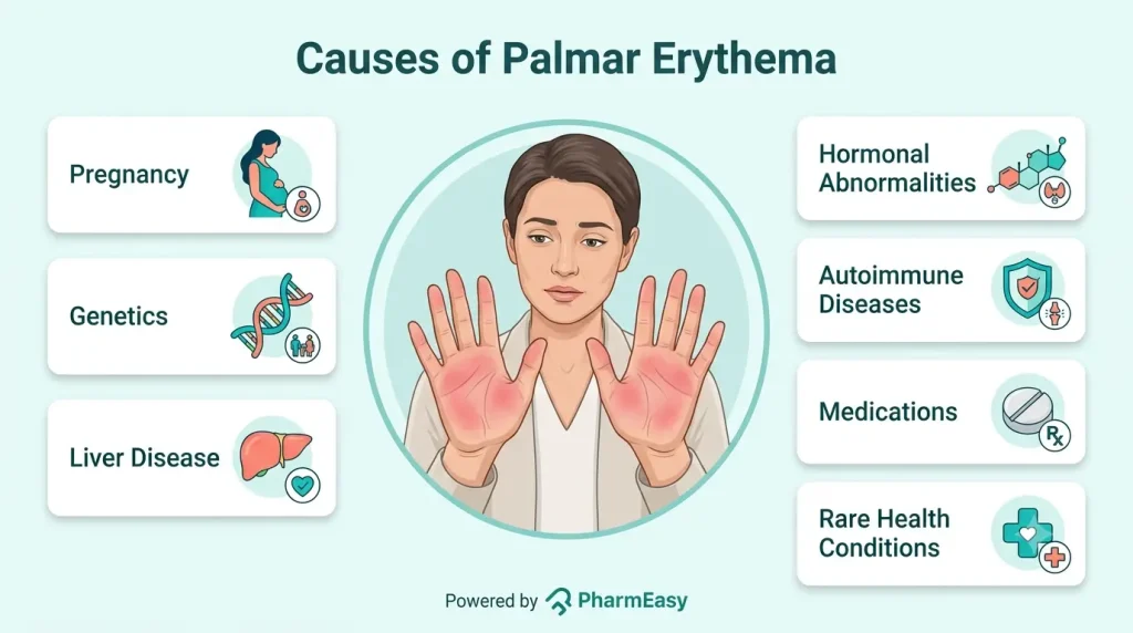
Palmar erythema may occur in several physiological and pathological conditions, including:
Palmar erythema is characterized by the following features:
Other associated symptoms depending upon the underlying cause such as autoimmune condition, liver disease, pregnancy etc, may also be noted along with the above-mentioned symptoms.
Some people are more likely to develop palmar erythema than others. People who are at a higher risk of developing palmar erythema include the following:
Palmar erythema is primarily a clinical diagnosis. Once identified, evaluation should focus on determining whether an underlying systemic disorder is present. Following the clinical examination, the primary cause of palmar erythema will be evaluated by the following:
The specific palmar erythema, rather the treatment plan depends on the underlying cause:
Palmar erythema often improves or resolves once the underlying cause is effectively treated. However, it may persist in chronic irreversible conditions such as liver cirrhosis4.
Also, the hereditary form of palmar erythema often persists throughout life and does not require any treatment unless if presents along with any associated disease or symptoms2,4.
Prevention of Palmar Erythema involves4:
Also Read: Liver Fibrosis: What Is It, Causes, Symptoms & Treatment
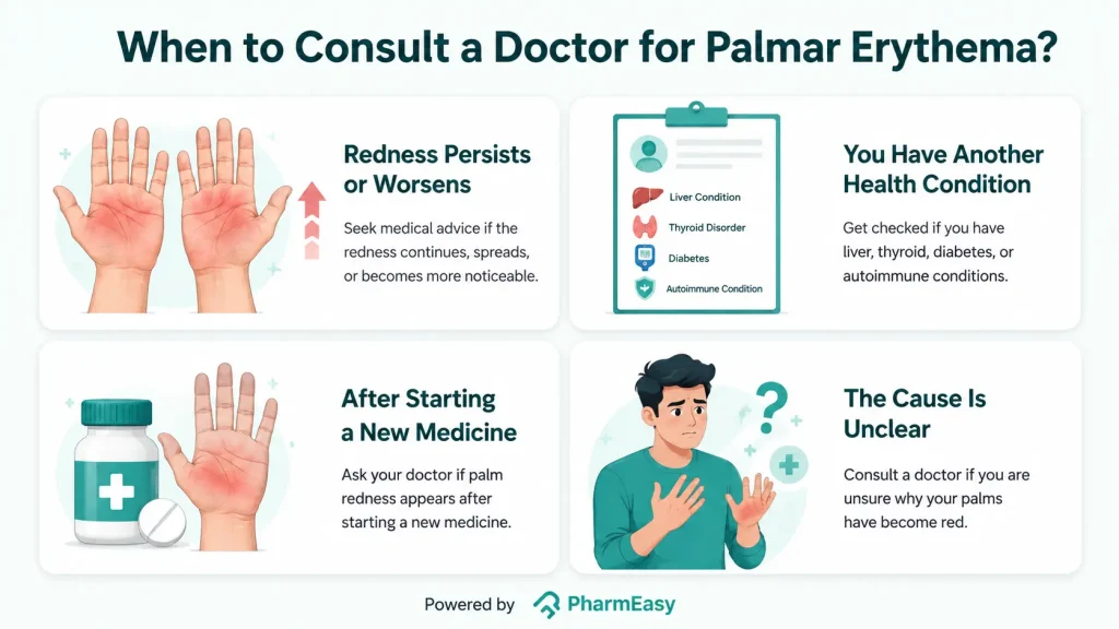
A doctor should be consulted for palmar erythema in the following conditions4:
Also Read: Facial Changes in Fatty Liver Disease: Causes, Diagnosis & Prevention
Palmar erythema is a condition in which redness of the palms occurs. Although palmar erythema is often benign, it may also serve as an important clinical sign of an underlying systemic disorder, particularly chronic liver disease. Persistent or unexplained palmar erythema warrants medical evaluation so that any associated condition can be identified and managed appropriately.
A liver that is not functioning properly leads to abnormal metabolization of hormones like oestradiol and vasoactive substances like nitric oxide. These cause dilation of capillaries and lead to redness and warmth in palms (palmar erythema) and sometimes soles3.
Yes, liver disease is one of the several diseases that are involved in causing palmar erythema. Several other conditions such as hyperthyroidism, diabetes, autoimmune, infections etc. have been found to be associated with palmar erythema4.
No, palmar erythema can be managed by controlling the underlying disease or condition. In many cases, palmar erythema improves after the underlying condition is treated, although it may persist in chronic diseases such as cirrhosis4.
The course of palmar erythema depends on its underlying cause. It may improve when the associated condition is treated, whereas hereditary palmar erythema usually persists throughout life6.
No, redness in the palms from palmar erythema is not known to be itchy6. Consult the doctor for guidance if it is felt itchy as it may indicate another condition or worsening of underlying disease.
Stress is not recognised as an established cause of palmar erythema. The condition is more commonly associated with pregnancy, liver disease, thyroid disorders, diabetes and autoimmune diseases4.
Smoking is not considered a direct cause of palmar erythema. However, it may contribute indirectly through its association with chronic liver and pulmonary diseases, which themselves have been linked to palmar erythema4˒11.
Menopause is not an established cause of palmar erythema. Hormonal conditions more commonly associated with palmar erythema include pregnancy, hyperthyroidism and diabetes4.
1. Bhandari A, Mahajan R. Skin Changes in Cirrhosis. J Clin Exp Hepatol. 2022;12(4):1215-1224. doi:10.1016/j.jceh.2021.12.013 https://pubmed.ncbi.nlm.nih.gov/35814509/
2. Langenauer J. Erythema Palmare Hereditarium (‘Red Palms’, ‘Lane’s Disease’). Case Rep Dermatol. 2014;6(3):245-247. doi:10.1159/000368822 https://pmc.ncbi.nlm.nih.gov/articles/PMC4255997/
3. Liu Y, Zhao Y, Gao X, et al. Recognizing skin conditions in patients with cirrhosis: a narrative review. Ann Med. 54(1):3017-3029. doi:10.1080/07853890.2022.2138961 https://pubmed.ncbi.nlm.nih.gov/36308406/
4. Serrao R, Zirwas M, English JC. Palmar erythema. Am J Clin Dermatol. 2007;8(6):347-356. doi:10.2165/00128071-200708060-00004 https://pubmed.ncbi.nlm.nih.gov/18039017/
5. Gurioli C, Patrizi A, Lambertini M, Neri I. Lane’s Disease (Erythema Palmare Hereditarium): A Report of Five Cases and a Review of the Literature. Pediatr Dermatol. 2017;34(5):590-594. doi:10.1111/pde.13211 https://pubmed.ncbi.nlm.nih.gov/28804916/
6. Palmar erythema. DermNet®. October 26, 2023. Accessed July 14, 2026. https://dermnetnz.org/topics/palmar-erythema
7. Lu J, Liu C, Zhou X, et al. Palmar erythema and palmar papules as predictors for dermatomyositis-related acute/subacute interstitial lung disease: a retrospective study. Rheumatology. 2021;61(1):413-421. doi:10.1093/rheumatology/keab177 https://pubmed.ncbi.nlm.nih.gov/33629112/
8. Noble JP, Boisnic S, Branchet-Gumila MC, Poisson M. Palmar erythema: cutaneous marker of neoplasms. Dermatology. 2002;204(3):209-213. doi:10.1159/000057883 https://europepmc.org/article/med/12037449
9. Palmar erythema and pruritus. Accessed July 14, 2026. https://www.aaaai.org/allergist-resources/ask-the-expert/answers/2023/palmar
10. Sow Y, Wachuku C, Barrera T, et al. Palmar and plantar erythema: An initial presentation of undifferentiated connective tissue disease. JAAD Case Rep. 2023;35:94-97. doi:10.1016/j.jdcr.2023.03.007 https://pmc.ncbi.nlm.nih.gov/articles/PMC10147949/
11. Pesut DP, Samardzic AM, Bulajic MV, Cvok-Debeljak TT. Palmar telangiectasia is associated with the intensity of smoking. J Bras Pneumol. 2019;45(2):e20180273. Published 2019 Apr 18. doi:10.1590/1806-3713/e20180273 https://pmc.ncbi.nlm.nih.gov/articles/PMC6733739/
12. Edwards E, Yosipovitch G. Skin Manifestations of Diabetes Mellitus. [Updated 2025 Mar 21]. In: Feingold KR, Adler RA, Ahmed SF, et al., editors. Endotext [Internet]. South Dartmouth (MA): MDText.com, Inc.; 2000-. Available from: https://www.ncbi.nlm.nih.gov/books/NBK481900/#
13. Kakroo SN, Banday S, Kakroo B, Beg MA. Cutaneous Manifestations of Respiratory Diseases. International Journal of Pharmaceutical and Clinical Research 2024; 16(9); 905-913. https://impactfactor.org/PDF/IJPCR/16/IJPCR,Vol16,Issue9,Article148.pdf
Disclaimer: The information provided here is for educational/awareness purposes only and is not intended to be a substitute for medical treatment by a healthcare professional and should not be relied upon to diagnose or treat any medical condition. The reader should consult a registered medical practitioner to determine the appropriateness of the information and before consuming any medication. PharmEasy does not provide any guarantee or warranty (express or implied) regarding the accuracy, adequacy, completeness, legality, reliability or usefulness of the information; and disclaims any liability arising thereof.
Links and product recommendations in the information provided here are advertisements of third-party products available on the website. PharmEasy does not make any representation on the accuracy or suitability of such products/services. Advertisements do not influence the editorial decisions or content. The information in this blog is subject to change without notice. The authors and administrators reserve the right to modify, add, or remove content without notification. It is your responsibility to review this disclaimer regularly for any changes.
Have you ever had ear pain that made it difficult to sleep, or noticed that sounds suddenly seem muffled? For parents, it can be especially worrying when a child wakes up at night crying and holding their ear, complaining of discomfort. Otitis media is an infection or inflammation of the middle ear. Fluid or mucus can build up behind the eardrum, which may cause ear pain, pressure, or muffled hearing1.
This blog explains what otitis media means, the main types of middle ear infection, common causes, symptoms, prevention tips, when to seek medical care, and how ear infections are usually treated.
An ear infection, also called otitis media when it affects the middle ear, often develops after a cold, sore throat, or other respiratory infection2. Fluid can collect behind the eardrum and make it harder for sound to pass through clearly1. Ear infections are more common in children, although adults can also develop them. A child with an ear infection may pull at the ear, cry more than usual, sleep poorly, have balance problems, or seem less responsive to sounds1.
There are three main types of middle ear infection. They differ mainly in how quickly symptoms start and how long fluid or infection remains behind the eardrum2.
A middle ear infection can be caused by bacteria or viruses. It often follows a cold or another upper respiratory infection, which can make the Eustachian tube swollen or blocked. This tube normally helps drain fluid from the middle ear and balance pressure3.
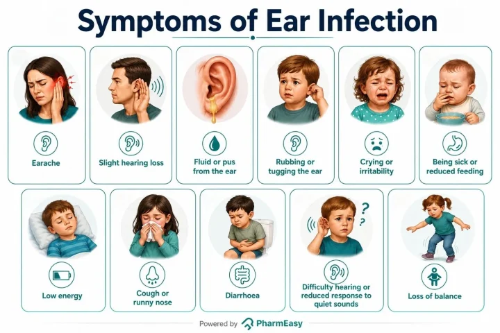
Middle ear infection symptoms often appear quickly and may improve over a few days. Symptoms can look different in adults, older children, babies, and younger children4.
Treatment depends on the person’s age, symptoms, general health, and risk of complications. Many middle ear infections improve on their own within a few days, so treatment may focus first on relieving pain and fever. Antibiotics are used when symptoms are severe, when the infection does not improve, or when the person is at higher risk of complications2,5.
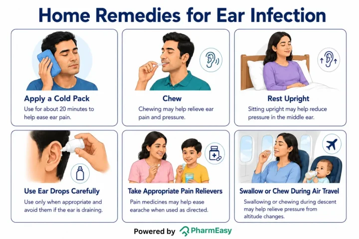
The following home-care steps may help ease earache or pressure while you wait for the infection to improve or while following medical advice6:
Most ear infections get better within a few days, and complications of otitis media are uncommon. However, when complications occur, the infection can spread to nearby parts of the ear or head4,7.
These steps may help lower the risk of middle ear infections:
Seek medical advice if you or your child has symptoms of an ear infection:
Also Read: Ear Bleeding (Otorrhagia): Causes, Symptoms, Treatment and Prevention
Middle ear infections, or otitis media, are common, particularly in children, and often improve on their own. Recognising symptoms such as ear pain, fever, hearing difficulties, or ear discharge can help you know when medical advice may be needed. Simple preventive measures and appropriate home care can help reduce discomfort and the risk of infection, while timely medical attention can help prevent complications.
Also Read: Natural Home Remedies for Ear Pain
Middle ear infections are not contagious. However, the cold or respiratory virus that may lead to an ear infection can spread from person to person8.
Yes. Otitis media can cause temporary hearing difficulty, especially when fluid remains in the middle ear. Permanent hearing loss is uncommon but can occur if infections are severe, repeated, or not treated when treatment is needed.
Most cases of otitis media improve within 3 to 5 days. Seek medical advice if symptoms do not improve within 2 to 3 days, get worse, or are accompanied by concerning symptoms such as high fever, ear discharge, or hearing loss7.
An ear infection may be serious if there is a high fever, worsening pain, swelling or redness behind the ear, fluid or pus draining from the ear, new hearing loss, dizziness, repeated vomiting, severe headache, neck stiffness, unusual drowsiness, breathing problems, dehydration, a seizure, or a rash that does not fade when pressed. Seek urgent medical help if any of these symptoms occur11.
Disclaimer: The information provided here is for educational/awareness purposes only and is not intended to be a substitute for medical treatment by a healthcare professional and should not be relied upon to diagnose or treat any medical condition. The reader should consult a registered medical practitioner to determine the appropriateness of the information and before consuming any medication. PharmEasy does not provide any guarantee or warranty (express or implied) regarding the accuracy, adequacy, completeness, legality, reliability or usefulness of the information; and disclaims any liability arising thereof.
Links and product recommendations in the information provided here are advertisements of third-party products available on the website. PharmEasy does not make any representation on the accuracy or suitability of such products/services. Advertisements do not influence the editorial decisions or content. The information in this blog is subject to change without notice. The authors and administrators reserve the right to modify, add, or remove content without notification. It is your responsibility to review this disclaimer regularly for any changes.
There is often a confusion between cholesterol and triglycerides, as both are types of lipids found in the blood. Elevated levels of either can significantly increase the risk of cardiovascular disease (CVD)1.
In 2022, an estimated 19.8 million people died from CVDs worldwide, accounting for approximately 32% of all global deaths, with heart attack and stroke responsible for 85% of these deaths2.
This blog explores what they are, how they differ, their ideal levels, and practical ways to keep them under control.
Cholesterol is a waxy, fat-like substance found in all cells of the body3. It plays an important role in building and maintaining cell walls and is needed to make hormones, vitamin D, and bile acids (substances that help to digest food). The liver makes most of the cholesterol the body needs, while the rest comes from foods such as meat, eggs, and dairy products. Although HDL cholesterol (good cholesterol) is essential for good health, too much LDL cholesterol (bad cholesterol) in the blood is a well-established cause of atherosclerotic cardiovascular disease (ASCVD)1,4. HDL participates in reverse cholesterol transport, but this mechanism is more complex than simply removing cholesterol from arteries.
Triglycerides are the most common type of fat in the body and are the body’s main way of storing energy5. After you eat, excess calories that the body does not need right away are converted into triglycerides and stored in fat cells for future energy use. When the body needs energy between meals or during physical activity, these stored triglycerides are broken down and used as fuel. Triglycerides also help keep the body warm and protect organs. However, high triglyceride levels are associated with an increased risk of heart disease. This risk is mainly linked to triglyceride-rich lipoproteins and remnant cholesterol (cholesterol carried in triglyceride-rich fat particles in your blood), rather than triglycerides directly causing the hardening and narrowing of the arteries5,6.
Note: Cholesterol helps build and maintain the body, while triglycerides store and provide energy. Both are necessary for normal functioning of the body; however, keeping their levels within a healthy range is important for protecting the heart.
The table below explains key details about triglycerides vs cholesterol to help you understand their roles better1,3,5,7:
| Feature | Cholesterol | Triglycerides |
| Main Function | Helps build cell membranes and is used to produce hormones, vitamin D, and bile acids for digestion | Stores excess calories and provides energy when the body needs it. |
| Primary Source | Produced mainly by the liver | Formed from excess calories consumed through food, especially carbohydrates, fats, and sugars |
| How It Travels in Blood | Carried by lipoproteins such as low-density lipoprotein (LDL) and high-density lipoprotein (HDL) | Transported primarily by chylomicrons and VLDL (very-low-density lipoproteins) |
| Normal Range | Total cholesterol: <200 mg/dL LDL: <100 mg/dL HDL: ≥60 mg/dL | <150 mg/dL |
| Health Risk When Elevated | High LDL cholesterol can accumulate on the inside of artery walls, increasing the risk of atherosclerosis, heart attack, and stroke | High triglyceride levels are associated with an increased risk of ASCVD (due to triglyceride-rich lipoproteins and remnant cholesterol). Very high triglyceride levels (≥500 mg/dL), especially ≥1000 mg/dL, can also greatly increase the risk of acute pancreatitis. |
The most common test used to measure cholesterol and triglycerides is lipid profile (lipid panel). This cholesterol and triglycerides test helps assess cardiovascular health and identify abnormalities in blood lipid levels8.
1. Purpose
2. What It Measures
3. Fasting Requirements
4. When to Get Tested
Note: Follow-up testing may be required to monitor response to treatment or lifestyle modifications.
Making healthy food choices may help improve cholesterol and triglyceride levels and reduce the risk of heart disease. Foods that can help lower cholesterol and triglyceride levels include9,10:
Foods that may increase cholesterol or triglyceride levels should be limited. These include:
Healthy lifestyle changes may help lower cholesterol and triglyceride levels and improve overall heart health. Some options include:



Also Read: High LDL (Bad) Cholesterol: Causes, Symptoms & How to Reduce It
While mild changes in cholesterol and triglyceride levels can often be managed through lifestyle modifications, it is important to consult a doctor if you have concerns about your heart health or lipid levels.
You should seek medical advice if:
Also Read: Low HDL (Good) Cholesterol: Causes, Symptoms & How to Increase It
Cholesterol and triglycerides are both essential fats that play important roles in the body; however, elevated levels of both can increase the risk of heart disease, heart attack, and stroke. Therefore, understanding the differences between cholesterol and triglycerides, knowing the levels of both, and getting regular lipid profile tests can help you stay on top of your cardiovascular health.
Healthy lifestyle habits (such as eating a balanced diet, staying physically active, maintaining a healthy weight, avoiding smoking, and limiting alcohol intake) can go a long way in improving cholesterol and triglyceride levels and reducing the risk of heart conditions.
Neither is necessarily ‘worse’ than the other. Both may increase the risk of CVDs when their levels are elevated. High LDL (bad) cholesterol is a well-established risk factor for atherosclerosis, while high triglycerides are also linked to heart disease and often occur alongside conditions such as obesity or diabetes3,5. However, your overall cardiovascular risk is determined by your complete lipid profile and other health conditions.
High triglycerides may occur even when cholesterol levels are normal. Common causes include consuming excess calories, diets high in sugar or refined carbohydrates, obesity, physical inactivity, excessive alcohol intake, uncontrolled diabetes, and certain medications5. Consulting a doctor can help identify the reason for elevated triglyceride levels and guide appropriate management.
Cholesterol and triglycerides are both lipids (serving different functions) that circulate in the bloodstream. Cholesterol helps build cells and produce hormones, while triglycerides store energy1.1 Both are measured in the lipid profile test8.
High cholesterol and triglycerides usually do not cause noticeable symptoms, including fatigue. However, they can contribute to CVDs that may result in fatigue, shortness of breath, or difficulty performing physical activities13. If you experience persistent fatigue, you should consult a doctor for further evaluation.
Triglycerides do not directly cause high cholesterol. However, both can be elevated due to similar factors such as an unhealthy diet, obesity, lack of physical activity, diabetes, or genetic conditions. As a result, high triglycerides and high cholesterol often occur together3,5.
Long-term stress may contribute to higher cholesterol and triglyceride levels. Stress triggers the release of hormones, which may affect how the body processes fats and can lead to increased triglyceride levels14. In addition, stress may lead to unhealthy lifestyle habits such as overeating, smoking, excessive alcohol consumption, and physical inactivity, which could contribute to elevated cholesterol and triglyceride levels12.
1. Cox RA, García-Palmieri MR. Cholesterol, Triglycerides, and Associated Lipoproteins. In: Walker HK, Hall WD, Hurst JW, eds. Clinical Methods: The History, Physical, and Laboratory Examinations. 3rd ed. Butterworths; 1990. Accessed June 2, 2026. http://www.ncbi.nlm.nih.gov/books/NBK351/
2. Cardiovascular diseases (CVDs). Accessed June 2, 2026. https://www.who.int/news-room/fact-sheets/detail/cardiovascular-diseases-(cvds)
3. Cholesterol. Accessed June 2, 2026. https://medlineplus.gov/cholesterol.html
4. Huff T, Boyd B, Jialal I. Physiology, Cholesterol. In: StatPearls. StatPearls Publishing; 2026. Accessed June 2, 2026. http://www.ncbi.nlm.nih.gov/books/NBK470561/
5. Triglycerides. Accessed June 2, 2026. https://medlineplus.gov/triglycerides.html
6. Services D of H& H. Triglycerides. Accessed June 2, 2026. http://www.betterhealth.vic.gov.au/health/conditionsandtreatments/triglycerides
7. Cholesterol Levels: What You Need to Know. Accessed June 2, 2026. https://medlineplus.gov/cholesterollevelswhatyouneedtoknow.html
8. Lipid profile test: MedlinePlus Medical Encyclopedia. Accessed June 2, 2026. https://medlineplus.gov/ency/article/007812.htm
9. How to Lower Cholesterol with Diet. Accessed June 2, 2026. https://medlineplus.gov/howtolowercholesterolwithdiet.html
10. Services D of H& H. Cholesterol – healthy eating tips. Accessed June 2, 2026. http://www.betterhealth.vic.gov.au/health/conditionsandtreatments/cholesterol-healthy-eating-tips
11. High cholesterol – How to lower your cholesterol. nhs.uk. May 28, 2019. Accessed June 2, 2026. https://www.nhs.uk/conditions/high-cholesterol/how-to-lower-your-cholesterol/
12. Jan 2 LR, 2025. Lifestyle Changes to Prevent a Heart Attack. www.heart.org. Accessed June 2, 2026. https://www.heart.org/en/health-topics/heart-attack/life-after-a-heart-attack/lifestyle-changes-for-heart-attack-prevention
13. Khan MR, Haider ZM, Hussain J, Malik FH, Talib I, Abdullah S. Comprehensive Analysis of Cardiovascular Diseases: Symptoms, Diagnosis, and AI Innovations. Bioengineering. 2024;11(12):1239. doi:10.3390/bioengineering11121239 https://pubmed.ncbi.nlm.nih.gov/39768057/
14. Anni NS, Jung SJ, Shim JS, Jeon YW, Lee GB, Kim HC. Stressful life events and serum triglyceride levels: the Cardiovascular and Metabolic Diseases Etiology Research Center cohort in Korea. Epidemiol Health. 2021;43:e2021042. doi:10.4178/epih.e2021042 https://pubmed.ncbi.nlm.nih.gov/34126706/
Disclaimer: The information provided here is for educational/awareness purposes only and is not intended to be a substitute for medical treatment by a healthcare professional and should not be relied upon to diagnose or treat any medical condition. The reader should consult a registered medical practitioner to determine the appropriateness of the information and before consuming any medication. PharmEasy does not provide any guarantee or warranty (express or implied) regarding the accuracy, adequacy, completeness, legality, reliability or usefulness of the information; and disclaims any liability arising thereof.
Links and product recommendations in the information provided here are advertisements of third-party products available on the website. PharmEasy does not make any representation on the accuracy or suitability of such products/services. Advertisements do not influence the editorial decisions or content. The information in this blog is subject to change without notice. The authors and administrators reserve the right to modify, add, or remove content without notification. It is your responsibility to review this disclaimer regularly for any changes.
Have you noticed more hair than usual on your pillow, in the shower, or on your comb? Hair loss is common. The most common type is androgenetic alopecia, also called genetic or pattern hair loss. It can affect both men (about 8 out of 10) and women (about 4 out of 10), although the pattern may look different1. Genes and hormones both play a role. One hormone involved is dihydrotestosterone (DHT)1. This article explains what DHT is, how it can affect hair growth, how doctors may assess DHT-related hair loss, and what lifestyle steps may support hair health.
DHT is a natural hormone made from testosterone with the help of an enzyme called 5-alpha-reductase. DHT assists normal male development, especially during puberty. It contributes to changes such as facial and body hair growth, a deeper voice, and growth of the prostate gland2. In adult women, however, DHT is not very beneficial2.
Did You Know?
Hair grows from tiny openings in the skin called hair follicles4. In people who are genetically sensitive to DHT, the hormone can attach to these follicles, especially on the scalp. Over time, the follicles may shrink. This process is called miniaturisation. Smaller follicles produce thinner and shorter hairs. The active growth phase may also become shorter, while the resting phase becomes longer. As this continues, some follicles may stop producing visible hair. Early diagnosis and treatment can help slow this process in some people1.
High DHT levels do not always cause noticeable symptoms. However, increased DHT activity may lead to certain changes in the body, especially in people who are genetically sensitive to its effects. Some possible symptoms of high DHT include:
Note: These symptoms may also be linked to other health conditions, so consulting a doctor is important for an accurate diagnosis.
Doctors do not usually diagnose hair loss by checking DHT alone. They first look at the pattern of hair loss, medical history, family history, scalp health, medicines, nutrition, and any symptoms that may suggest a hormone or thyroid problem.
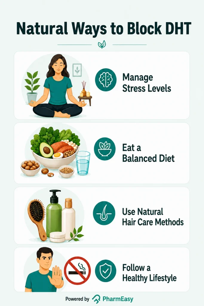
Many people search for ways to reduce DHT when they learn that DHT is linked to pattern hair loss. Lifestyle changes can support general hair and scalp health, but they may not directly lower DHT enough to treat androgenetic alopecia. Medical treatments may be considered by a doctor when appropriate.
Note: The results of these approaches may differ depending on the individual. If you are experiencing persistent hair loss or noticeable hair thinning, it is best to consult a doctor for proper evaluation and treatment.
Some foods contain nutrients or plant compounds that have been studied for possible effects on hormone pathways linked to DHT. However, most evidence is early, indirect, or based on small studies. These foods may support general health, but they should not be described as proven DHT blockers.
Note: Including these foods as part of a balanced diet may support overall health, but they should not be an alternative for medical treatment. If you have persistent hair loss or concerns about high DHT levels, consult your doctor for proper evaluation and guidance.
Several supplements are marketed as “DHT blockers.” The table below explains the difference between food-based approaches and supplements.
| Natural DHT Blockers (Food Sources) | DHT Blocker Supplements |
| Whole foods that contain different nutrients such as vitamins, minerals, antioxidants, and healthy fats along with possible DHT-related benefits10,12,15. | Contain concentrated amounts of specific ingredients, such as from saw palmetto (a type of palm), that are marketed for their likely DHT-blocking effects16. |
| Can be used as part of a healthy eating pattern, which can support overall wellness. | Effectiveness may vary depending on the ingredients they contain17,18. |
| Usually safe for most people when eaten as part of a balanced diet, unless a person has an allergy or specific dietary restriction. | May carry a risk of side effects or interactions, especially when taken in high doses or with certain medications17. |
| May support hair health through nutrients and antioxidants, but any direct effect on DHT is uncertain10–14. | Some supplements may aim to reduce the conversion of testosterone to DHT, but strength, quality, safety, and results may vary17. |
Note: Foods and supplements should not replace medical treatment. Supplements can interact with medicines and may not be suitable for everyone. Prescription medicines are approved for specific uses, but over-the-counter “DHT blocker” supplements are not approved by the FDA to treat hair loss. Speak with a qualified doctor before using any supplement or medicine for hair loss.
Medicines that lower DHT can cause side effects in some people. These effects are more commonly discussed in men using prescription DHT-lowering medicines, but anyone considering treatment should review benefits and risks with a doctor.
Consult a doctor if your hair fall becomes persistent, worsens over time, or starts affecting your daily life. Medical advice is also important if you notice sudden hair shedding, a receding hairline, thinning hair, or bald patches1,2. If your scalp feels itchy, painful, red, or flaky along with hair loss, it could indicate an underlying scalp condition that requires treatment19.
If you are taking DHT-blocking medications or supplements and develop side effects such as reduced sex drive, erectile or ejaculatory problems, skin rashes, mood changes, or breast enlargement (in men), consult your doctor promptly17,18.
Hair loss that begins after starting a new medication, recovering from a recent illness, or experiencing significant stress should also be evaluated20. In women with PCOS, symptoms such as hair fall, irregular periods, excessive facial hair, or severe acne may indicate a hormonal imbalance that requires medical attention6.
Also Read: Blood Test for Hair Loss: Who Should Get Tested, Types and Prevention
Understanding DHT can help explain why pattern hair loss happens in some people. DHT is a normal hormone, but people who are genetically sensitive to it may develop gradual hair thinning. Healthy habits, gentle hair care, and good nutrition can support hair health, but persistent or worsening hair loss should be assessed by a doctor. Early medical advice can help identify the cause and discuss treatment options that may slow hair loss or improve hair growth.
Lowering DHT may help slow hair loss and protect existing hair follicles in some people with androgenetic alopecia. Regrowth is possible for some people, especially when treatment starts early, but results vary. A doctor can help decide whether prescription-based treatments are suitable18.
You usually cannot tell that DHT is high from symptoms alone. A doctor may look at your hair loss pattern, scalp, medical history, family history, medicines, and symptoms such as acne, excess facial or body hair, irregular periods, or prostate-related symptoms. A DHT blood test may be used in selected cases, but it is not the only way to assess hair loss1,3.
DHT-related hair loss can often be slowed, and some people may see thicker hair with the right treatment. Complete reversal is less likely when follicles have stopped producing visible hair for a long time. Early medical advice gives the best chance of preserving existing hair18.
DHT may make oil glands in the skin more active. This can lead to oily skin and may contribute to acne in some people, but acne can have many causes. If acne is persistent, painful, or causing scarring, it is best to speak with a doctor or dermatologist21.
Current evidence is not enough to confirm that creatine raises DHT or directly causes DHT-related hair loss. If you notice hair shedding after starting any supplement, stop guessing and speak with a healthcare professional, especially if hair loss is sudden or worsening22.
Some prescription medicines that lower DHT may cause erectile problems in some people, although this does not happen to everyone. Do not stop a prescribed medicine without medical advice. If you notice sexual side effects, mood changes, breast tenderness, or any worrying symptom, contact your doctor18.
1. Bazargan AS, Jafarzadeh A, Ayoubi A, et al. Investigating the relationship between androgenetic alopecia and hair shape, color, and thickness: A case‐control study. Health Sci Rep. 2025;8(5):e70764. doi:10.1002/hsr2.70764. Available from: https://pmc.ncbi.nlm.nih.gov/articles/PMC12040711/
2. Kinter KJ, Amraei R, Anekar AA. Biochemistry, dihydrotestosterone. In: StatPearls. StatPearls Publishing; 2026. Accessed August 6, 2026. Available from: http://www.ncbi.nlm.nih.gov/books/NBK557634/
3. Swerdloff RS, Dudley RE, Page ST, Wang C, Salameh WA. Dihydrotestosterone: Biochemistry, Physiology, and Clinical Implications of Elevated Blood Levels. Endocr Rev. 2017;38(3):220-254. doi:10.1210/er.2016-1067. Available from: https://pubmed.ncbi.nlm.nih.gov/28472278/
4. Hair. Better Health Channel. 2023. Accessed August 6, 2026. Available from: https://www.betterhealth.vic.gov.au/health/conditionsandtreatments/hair
5. Makrantonaki E, Ganceviciene R, Zouboulis C. An update on the role of the sebaceous gland in the pathogenesis of acne. Dermatoendocrinology. 2011;3(1):41-49. doi:10.4161/derm.3.1.13900. Available from: https://pmc.ncbi.nlm.nih.gov/articles/PMC3051853/
6. Ebrahimian A, Moradi A, Kheyri V, Najafipour F, Sadra V. Evaluation of dihydrotestosterone levels and total testosterone to dihydrotestosterone ratio with clinical symptoms and metabolic parameters in patients with polycystic ovary syndrome. BMC Endocr Disord. 2025;25:263. doi:10.1186/s12902-025-02070-4. Available from: https://pmc.ncbi.nlm.nih.gov/articles/PMC12617002/
7. Gokce N, Basgoz N, Kenanoglu S, et al. An overview of the genetic aspects of hair loss and its connection with nutrition. J Prev Med Hyg. 2022;63(2 Suppl 3):E228-E238. doi:10.15167/2421-4248/jpmh2022.63.2S3.2765. Available from: https://pmc.ncbi.nlm.nih.gov/articles/PMC9710406/
8. Relaxation techniques – relieving stress and anxiety. Healthdirect. 2026. Accessed August 6, 2026. Available from: https://www.healthdirect.gov.au/relaxation-techniques-for-stress-relief
9. Hair styling without damage. Accessed August 6, 2026. Available from: https://www.aad.org/public/diseases/hair-loss/hair-care/styling
10. Dhamija P, More A, Choudhary N, Wadhe T, Barai J, Shah D. Seed Cycling and Hormonal Balance: A Case Study of Successful Fertility Intervention in Polycystic Ovarian Syndrome. J Pharm Bioallied Sci. 2025;17(Suppl 1):S1030-S1033. doi:10.4103/jpbs.jpbs_164_25. Available from: https://pmc.ncbi.nlm.nih.gov/articles/PMC12156535/
11. Naftalin RJ, Afzal I, Cunningham P, et al. Interactions of androgens, green tea catechins and the antiandrogen flutamide with the external glucose-binding site of the human erythrocyte glucose transporter GLUT1. Br J Pharmacol. 2003;140(3):487-499. doi:10.1038/sj.bjp.0705460. Available from: https://pmc.ncbi.nlm.nih.gov/articles/PMC1574051/
12. Arias-Chávez DJ, Mailloux-Salinas P, Ledesma-Aparicio J, et al. Tomato lipidic extract plus selenium decrease prostatic hyperplasia, dihydrotestosterone and androgen receptor expression versus finasteride in rats. World J Urol. 2023;41(10):2793-2799. doi:10.1007/s00345-023-04558-x. Available from: https://pubmed.ncbi.nlm.nih.gov/37659980/
13. Dillingham BL, McVeigh BL, Lampe JW, Duncan AM. Soy protein isolates of varying isoflavone content exert minor effects on serum reproductive hormones in healthy young men. J Nutr. 2005;135(3):584-591. doi:10.1093/jn/135.3.584. Available from: https://pubmed.ncbi.nlm.nih.gov/15735098/
14. Ide H, Lu Y, Noguchi T, et al. Modulation of AKR1C2 by curcumin decreases testosterone production in prostate cancer. Cancer Sci. 2018;109(4):1230-1238. doi:10.1111/cas.13517. Available from: https://pubmed.ncbi.nlm.nih.gov/29369461/
15. Raza N, Sadaf A, Mushtaq R, et al. Nutritional and Health Potential of Edible Seeds: Micronutrient Bioavailability and Mechanistic Insights. Food Sci Nutr. 2026;14(2):e71480. doi:10.1002/fsn3.71480. Available from: https://pmc.ncbi.nlm.nih.gov/articles/PMC12852966/
16. Evron E, Juhasz M, Babadjouni A, Mesinkovska NA. Natural Hair Supplement: Friend or Foe? Saw Palmetto, a Systematic Review in Alopecia. Skin Appendage Disord. 2020;6(6):329-337. doi:10.1159/000509905. Available from: https://pmc.ncbi.nlm.nih.gov/articles/PMC7706486/
17. Ashique S, Sandhu NK, Haque SkN, Koley K. A Systemic Review on Topical Marketed Formulations, Natural Products, and Oral Supplements to Prevent Androgenic Alopecia: A Review. Nat Prod Bioprospect. 2020;10(6):345-365. doi:10.1007/s13659-020-00267-9. Available from: https://pmc.ncbi.nlm.nih.gov/articles/PMC7648777/
18. Devjani S, Ezemma O, Kelley KJ, Stratton E, Senna M. Androgenetic Alopecia: Therapy Update. Drugs. 2023;83(8):701-715. doi:10.1007/s40265-023-01880-x. Also Read: https://pmc.ncbi.nlm.nih.gov/articles/PMC10173235/
19. Dandruff, Cradle Cap, and Other Scalp Conditions: MedlinePlus. Accessed August 6, 2026. https://medlineplus.gov/dandruffcradlecapandotherscalpconditions.html
20. Singh S, Muthuvel K. Practical Approach to Hair Loss Diagnosis. Indian J Plast Surg. 2021;54(4):399-403. doi:10.1055/s-0041-1739240. Available from: https://pmc.ncbi.nlm.nih.gov/articles/PMC8719967/
21. Lee WJ, Jung HD, Chi SG, et al. Effect of dihydrotestosterone on the upregulation of inflammatory cytokines in cultured sebocytes. Arch Dermatol Res. 2010;302(6):429-433. doi:10.1007/s00403-009-1019-6. Available from: https://www.researchgate.net/publication/40820124_Effect_of_dihydrotestosterone_on_the_upregulation_of_inflammatory_cytokines_in_cultured_sebocytes
22. Lak M, Forbes SC, Ashtary-Larky D, et al. Does creatine cause hair loss? A 12-week randomized controlled trial. J Int Soc Sports Nutr. 22(Suppl 1):2495229. doi:10.1080/15502783.2025.2495229. Available from: https://pmc.ncbi.nlm.nih.gov/articles/PMC12020143/
Disclaimer: The information provided here is for educational/awareness purposes only and is not intended to be a substitute for medical treatment by a healthcare professional and should not be relied upon to diagnose or treat any medical condition. The reader should consult a registered medical practitioner to determine the appropriateness of the information and before consuming any medication. PharmEasy does not provide any guarantee or warranty (express or implied) regarding the accuracy, adequacy, completeness, legality, reliability or usefulness of the information; and disclaims any liability arising thereof.
Links and product recommendations in the information provided here are advertisements of third-party products available on the website. PharmEasy does not make any representation on the accuracy or suitability of such products/services. Advertisements do not influence the editorial decisions or content. The information in this blog is subject to change without notice. The authors and administrators reserve the right to modify, add, or remove content without notification. It is your responsibility to review this disclaimer regularly for any changes.
Finding a high gamma-glutamyl transferase (GGT) level on a blood test can feel worrying, especially if you are not sure what it means. GGT is a liver-related enzyme that may rise when the liver or bile ducts are irritated or damaged. A high result does not diagnose a specific condition by itself, but it can help your doctor decide whether more tests are needed. This article explains what GGT is, what can raise it, possible symptoms and risks, treatment options, and when to seek medical advice.
GGT is an enzyme that is found in many tissues of the body, especially the liver and bile ducts. If liver or bile duct cells are damaged, GGT can leak into the blood, and the level may rise1.
Did you know?
The normal range for GGT can differ between laboratories and based on the assays. It can also vary based on the age or sex of the individual. Therefore, the reference range should not be representative of all laboratories, and priority should be given to the laboratory’s own reference range. Many reports list a range close to:
Your own laboratory report is the best guide because normal values can vary by age, sex, testing method, and laboratory. A result should be interpreted with your symptoms, medical history, medicines, alcohol use, and other liver test results3.
GGT can be widely distributed in the human body. However, high activity is seen in the liver, lining of bile ducts (biliary epithelium), kidney, pancreas, and some other tissues. Elevation of serum GGT is mainly seen in conditions affecting the liver and bile ducts. GGT elevation might not be a result of leakage from damaged cells in every case. Instead, it can also be due to reduced bile flow (cholestasis) and induction of GGT enzyme by certain drugs or alcohol intake. Detection of GGT level is commonly included in liver chemistry or liver enzyme panels; however, it cannot be considered a marker of liver function. Liver function is mainly represented by parameters such as albumin and International normalised ratio (INR), and excretory function of the liver by bilirubin.
There is no single GGT level that is “dangerous” for everyone. The level matters, but so do your symptoms, other liver tests, and medical history. In general, doctors look at how far the result is above the laboratory’s reference range:
A GGT test alone cannot identify the exact cause of a health problem. It is a clue that needs to be considered with other tests, such as alanine aminotransferase, aspartate aminotransferase, ALP, bilirubin, and sometimes imaging tests. Your healthcare provider can explain what your result means for you.
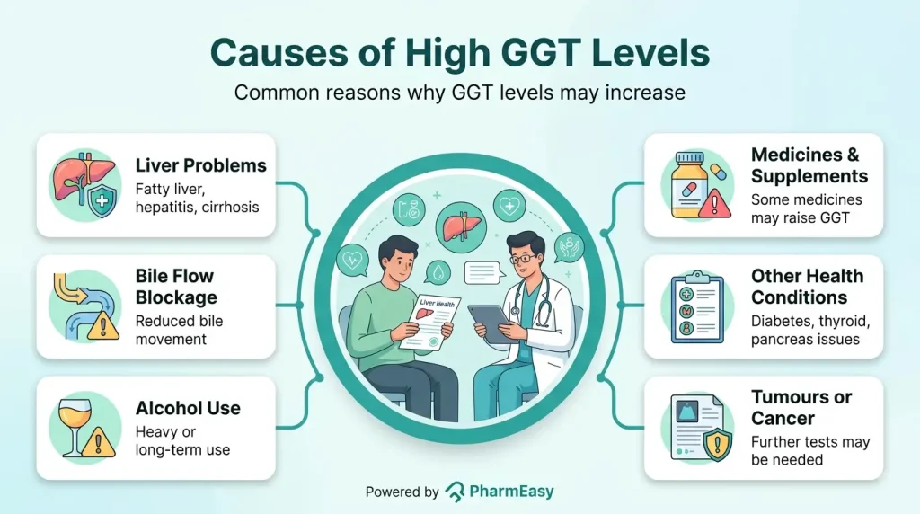
A high GGT level can be linked to liver or bile duct conditions, alcohol use, some medicines, and certain long-term health conditions. Common causes include6:
Elevated GGT can be seen associated with intake of alcohol, cholestasis, and certain drugs that are enzyme inducers. However, elevated GGT does not reflect the severity of liver damage.
High GGT itself usually does not cause symptoms. Symptoms, when present, come from the condition that is affecting the liver, bile ducts, or another organ. Possible symptoms include:
A persistently high GGT level may be linked with a higher chance of certain health problems, but GGT is usually a marker rather than the direct cause. It should be interpreted together with other test results and your overall health10.
GGT elevation is usually detected or measured from a test report rather than being diagnosed. The diagnosis focuses on the underlying cause that led to the elevation. GGT is usually checked with other liver tests. Doctors use the full pattern of results, your symptoms, your medical history, and sometimes imaging tests to understand what may be causing the high level11.
A single abnormal GGT result does not confirm a diagnosis. If your result is high, discuss it with your doctor, especially if you have symptoms or other abnormal liver tests.
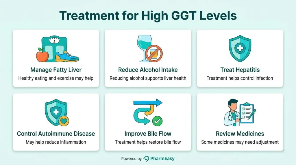
Treatment focuses on the cause of the high GGT level rather than the number itself. On the contrary, a decrease in GGT level after treatment may not mean that the treatment was successful. Your doctor may recommend one or more of the following, depending on the diagnosis13:
You may not be able to prevent every cause of high GGT, but these steps can support liver health and lower the risk of several related conditions:
If you drink, follow your doctor’s advice about safe limits. If you have liver disease, you may be advised to avoid alcohol completely. In advanced stages of liver disease, especially linked with the consumption of alcohol or liver cirrhosis or fibrosis, alcohol abstinence is advised instead of limiting the intake.

Gradual weight loss can reduce fat and inflammation in the liver for people with fatty liver disease14.
Choose fibre-rich foods, limit sugary drinks, and aim for regular physical activity as advised by your healthcare provider.

Tell your doctor about all medicines, supplements, and herbal products you take because some can affect the liver. However, the stoppage of medicines should not be based solely on GGT levels. Changes in medicines are only advised for those who might be associated with liver toxicity or enzyme induction. These changes can only be done under the supervision of a medical professional.

If you have cirrhosis, hepatitis, or another liver condition, regular monitoring helps detect changes early13.
Also Read: Liver Fibrosis: What Is It, Causes, Symptoms & Treatment
GGT is an enzyme found mainly in the liver and bile ducts. A high GGT level can suggest liver or bile duct irritation, but it does not diagnose a disease on its own. The most useful next step is to review the result with your doctor, who can compare it with your symptoms, medical history, medicines, alcohol use, and other test results. In many cases, treating the underlying cause and making liver-friendly lifestyle changes can help improve GGT levels over time.
If alcohol is the main cause, GGT may gradually fall after several weeks without alcohol15. If another condition is causing the rise, GGT may improve when that condition is treated.
There is no single alarming number for everyone. A result that is several times above the lab’s reference range, or a high result with symptoms such as jaundice, dark urine, pale stools, severe itching, or abdominal swelling, should be discussed promptly with a doctor4.
High GGT is not treated directly. It may return closer to normal when the cause is managed, such as reducing alcohol intake, treating hepatitis, improving fatty liver disease, or adjusting a medicine under medical supervision15.
No. GGT is an enzyme, not a disease. Fatty liver disease is one possible reason for a raised GGT, but other causes are also possible6.
Statins are the most common drugs that are used for the management of heart-related diseases. The common side effects associated with them are muscle dysfunction (myopathy) and elevation of some liver enzymes such as ALT or AST in the blood. However, there are rare cases of GGT elevation promoted by statins16.
Yes. Alcohol can increase GGT levels, especially with heavy or long-term drinking. If you are concerned about alcohol use, ask a healthcare professional for support15.
Dehydration is not a common cause of high GGT in people. High GGT is more often linked with liver disease, bile duct problems, alcohol use, certain medicines, or other health conditions17.
No. GGT can be seen as increased in many conditions, including liver cancers. However, it is not specific to cancer. It is mostly associated with liver diseases; however, it can also be seen in some other dysfunctions and alcoholism6.
Smoking has been linked with higher GGT levels in some studies. Smoking and alcohol together may have a stronger effect on liver-related test results18.
1. Kunutsor SK. Gamma-glutamyltransferase-friend or foe within? Liver Int Off J Int Assoc Study Liver. 2016;36(12):1723-1734. doi:10.1111/liv.13221 Available from: https://pubmed.ncbi.nlm.nih.gov/27512925/
2. Torrente MP, Freeman WM, Vrana KE. Protein biomarkers of alcohol abuse. Expert Rev Proteomics. 2012;9(4):425-436. doi:10.1586/epr.12.38 Available from: https://pubmed.ncbi.nlm.nih.gov/22967079/
3. Content – Health Encyclopedia – URochester Medicine. Accessed August 6, 2026. Available from: https://www.urmc.rochester.edu/encyclopedia/content?contenttypeid=167&contentid= gamma_glutamyl_transpeptidase
4. Vroon DH, Israili Z. Alkaline Phosphatase and Gamma Glutamyltransferase. In: Walker HK, Hall WD, Hurst JW, eds. Clinical Methods: The History, Physical, and Laboratory Examinations. 3rd ed. Butterworths; 1990. Accessed August 6, 2026. Available from: http://www.ncbi.nlm.nih.gov/books/NBK203/
5. Giannini EG, Testa R, Savarino V. Liver enzyme alteration: a guide for clinicians. CMAJ Can Med Assoc J. 2005;172(3):367-379. doi:10.1503/cmaj.1040752 Available from: https://pubmed.ncbi.nlm.nih.gov/15684121/
6. H.A. K. The Serum Gamma Glutamyl Transpeptidase – A Non invasive Diagnostic Bio Marker of Chronic Anicteric Non Alcoholic Liver Diseases. J Clin Diagn Res JCDR. 2013;7(4):691-694. doi:10.7860/JCDR/2013/5569.2883 Available from: https://pmc.ncbi.nlm.nih.gov/articles/PMC3644446/
7. Gamma-glutamyl Transferase (GGT) Test: MedlinePlus Medical Test. Accessed August 5, 2026. Available from: https://medlineplus.gov/lab-tests/gamma-glutamyl-transferase-ggt-test/
8. Symptoms & Causes of Cirrhosis – NIDDK. National Institute of Diabetes and Digestive and Kidney Diseases. Accessed August 5, 2026. Available from: https://www.niddk.nih.gov/health-information/liver-disease/cirrhosis/symptoms-causes
9. Cholestasis: MedlinePlus Medical Encyclopedia. Accessed August 5, 2026. Available from: https://medlineplus.gov/ency/article/000215.htm
10. Cho EJ, Jeong SM, Chung GE, et al. Gamma-glutamyl transferase and risk of all-cause and disease-specific mortality: a nationwide cohort study. Sci Rep. 2023;13:1751. doi:10.1038/s41598-022-25970-0 Available from: https://pubmed.ncbi.nlm.nih.gov/36720971/
11. Diagnosis of NAFLD & NASH – NIDDK. National Institute of Diabetes and Digestive and Kidney Diseases. Accessed August 5, 2026. Available from: https://www.niddk.nih.gov/health-information/liver-disease/nafld-nash/diagnosis
12. Liver Function Tests: MedlinePlus Medical Test. Accessed August 6, 2026. Available from: https://medlineplus.gov/lab-tests/liver-function-tests/
13. Treatment for Cirrhosis – NIDDK. National Institute of Diabetes and Digestive and Kidney Diseases. Accessed August 6, 2026. Available from: https://www.niddk.nih.gov/health-information/liver-disease/cirrhosis/treatment
14. Treatment for NAFLD & NASH – NIDDK. National Institute of Diabetes and Digestive and Kidney Diseases. Accessed August 6, 2026. Available from: https://www.niddk.nih.gov/health-information/liver-disease/nafld-nash/treatment
15. Horner F, Kellen JA, Kingstone E, Maharaj N, Malkin A. Dynamic changes of serum gamma-glutamyl transferase in chronic alcoholism. Enzyme. 1979;24(4):217-223. doi:10.1159/000458662 Available from: https://pubmed.ncbi.nlm.nih.gov/39748/
16. Xu Y, Wu Y. Atorvastatin associated with gamma glutamyl transpeptidase elevation in a hyperlipidemia patient: A case report and literature review. Medicine (Baltimore). 2020;99(40):e22572. doi:10.1097/MD.0000000000022572 Available from: https://pubmed.ncbi.nlm.nih.gov/33019469/
17. Sala G, Bronzo V, Boccardo A, et al. Assessing failure of transfer of passive immunity by gamma-glutamyl-transferase activity and serum refractometry in holstein-friesian calves affected by neonatal diarrhea. Vet Res Commun. 2023;47(4):2315-2321. doi:10.1007/s11259-023-10149-3 Available from: https://pubmed.ncbi.nlm.nih.gov/37314566/
18. Zhang Z, Ma L, Geng H, Bian Y. Effects of Smoking, and Drinking on Serum Gamma-Glutamyl Transferase Levels Using Physical Examination Data: A Cross-Sectional Study in Northwest China. Int J Gen Med. 2021;14:1301-1309. doi:10.2147/IJGM.S301900 Available from: https://pubmed.ncbi.nlm.nih.gov/33883928/
Disclaimer: The information provided here is for educational/awareness purposes only and is not intended to be a substitute for medical treatment by a healthcare professional and should not be relied upon to diagnose or treat any medical condition. The reader should consult a registered medical practitioner to determine the appropriateness of the information and before consuming any medication. PharmEasy does not provide any guarantee or warranty (express or implied) regarding the accuracy, adequacy, completeness, legality, reliability or usefulness of the information; and disclaims any liability arising thereof.
Links and product recommendations in the information provided here are advertisements of third-party products available on the website. PharmEasy does not make any representation on the accuracy or suitability of such products/services. Advertisements do not influence the editorial decisions or content. The information in this blog is subject to change without notice. The authors and administrators reserve the right to modify, add, or remove content without notification. It is your responsibility to review this disclaimer regularly for any changes.
Skin concerns, such as itchy rashes, unexplained skin irritation, or discomfort that keeps coming back, could be quite bothersome. Some skin conditions may be linked to underlying health issues and require proper evaluation.
Dermatitis herpetiformis is a long-term, very itchy skin condition that causes small blisters, bumps, and sore patches. It happens when the immune system reacts to gluten, and this reaction affects the skin1,2.
Understanding dermatitis herpetiformis causes, its symptoms, and the treatments involved may help individuals detect and manage this condition early and effectively.
Dermatitis herpetiformis, also as Duhring disease, is a long-lasting autoimmune skin condition. It usually causes an intensely itchy rash with small bumps or blisters on both sides of the limbs and body, especially on the elbows, knees, buttocks, scalp, and back3. Because the blisters are often scratched, a person may notice scabs, scratches, or sore areas instead of clear blisters1.
Dermatitis herpetiformis symptoms include,2:
Dermatitis herpetiformis causes:
Some individuals are more vulnerable to getting Dermatitis Herpetiformis than others. These include:
If untreated or poorly managed, dermatitis herpetiformis can progress into more serious conditions, as follows:
A doctor may diagnose dermatitis herpetiformis using the following tests and assessments:
Treatment for dermatitis herpetiformis usually focuses on a strict gluten-free diet for long-term control. Because gluten can be hidden in many foods. It is helpful to consult a dietitian before making major diet changes. The rash may take months or longer to improve after starting a gluten-free diet, so some people may also need medications for symptomatic relief1. Treatment may include:
A gluten-free diet removes foods that contain wheat, barley, and rye. Some people may also need to avoid oats unless they are certified gluten-free and approved by their healthcare team4.

Doctors may prescribe medicines such as dapsone or other options to reduce itching and blisters. These medicines need medical supervision and monitoring for side effects1.

Avoid exposure to iodine-containing foods/medications, as it can trigger dermatitis herpetiformis symptoms1.
Dermatitis herpetiformis does not have a known prevention. Prevention of the complications of the condition could include the following1:
Also Read: Typhoid Fever Rash: When Rose Spots Appear & What You Should Do
Dermatitis herpetiformis is a long-term skin condition linked to gluten sensitivity and coeliac disease. It can cause severe itching, small blisters, and sore or scratched skin. A strict gluten-free diet is the main long-term treatment, and medicines may help control symptoms while the diet takes effect. Anyone with a persistent, very itchy rash should speak with a healthcare professional for proper testing and treatment.
With the right diagnosis, medical follow-up, and diet support, many people can reduce flare-ups and lower the risk of related complications.
Dermatitis herpetiformis is associated with severe itching, scars, skin erosions, and a rash that is symmetrical on the back, buttocks, knees, and elbows. It is similar to the rash in eczema2.
Sensitivity to gluten and gliadin (a derivative of gluten) can cause the characteristic rash suggestive of dermatitis herpetiformis, which could be diagnosed through physical examination, blood tests, and a skin biopsy1,7.
Yes, it could occur at any time in life, depending on the extent of exposure to gluten, the immunity at the time, and the overall health of the individual. It is often seen in individuals who are 15 to 40 years old4,8.
No, dermatitis herpetiformis is not contagious. It is an autoimmune, lifelong condition that affects the skin and could be hereditary. It is not an infection that could spread from one person to another9.
Even though the name may suggest herpes, it has no relevance to the herpes virus, and it is an autoimmune condition where the immune cells fail to recognise their own cells2.
Excess alcohol intake could make an individual more sensitive to gluten, particularly if a close family relative is known to have dermatitis herpetiformis. In an individual already diagnosed with the condition, alcohol intake could worsen the symptoms1,10.
No, stress is not known to cause dermatitis herpetiformis. It is mainly caused by immune sensitivity to gluten1.
Yes, the rash in dermatitis herpetiformis may seem like acne because it also appears like a small, papular lesion. However, it also looks similar to an eczema rash. Therefore, simple visual diagnosis is challenging2.
1. Mirza HA, Gharbi A, Bhutta BS. Dermatitis Herpetiformis. In: StatPearls. StatPearls Publishing; 2026. Accessed July 28, 2026. Available from: http://www.ncbi.nlm.nih.gov/books/NBK493163/
2. Dermatitis herpetiformis: MedlinePlus Medical Encyclopedia. Accessed July 28, 2026. Available from: https://medlineplus.gov/ency/article/001480.htm
3. Nguyen CN, Kim SJ. Dermatitis Herpetiformis: An Update on Diagnosis, Disease Monitoring, and Management. Medicina (Mex). 2021;57(8):843. doi:10.3390/medicina57080843 Available from: https://pubmed.ncbi.nlm.nih.gov/34441049/
4. Dermatitis herpetiformis symptoms and treatments. NHS inform. Accessed July 28, 2026. Available from: https://www.nhsinform.scot/illnesses-and-conditions/skin/rashes-irritation-and-swelling/dermatitis-herpetiformis/
5. Reunala T, Hervonen K, Salmi T. Dermatitis Herpetiformis: An Update on Diagnosis and Management. Am J Clin Dermatol. 2021;22(3):329-338. doi:10.1007/s40257-020-00584-2 Available from: https://pubmed.ncbi.nlm.nih.gov/33432477/
6. Dermatitis Herpetiformis – NIDDK. National Institute of Diabetes and Digestive and Kidney Diseases. Accessed July 28, 2026. Available from: https://www.niddk.nih.gov/health-information/professionals/clinical-tools-patient-management/digestive-diseases/dermatitis-herpetiformis
7. Collin P, Salmi TT, Hervonen K, Kaukinen K, Reunala T. Dermatitis herpetiformis: a cutaneous manifestation of coeliac disease. Ann Med. 2017;49(1):23-31. doi:10.1080/07853890.2016.1222450 Available from: https://pubmed.ncbi.nlm.nih.gov/27499257/
8. Reunala T, Hervonen K, Salmi T. Dermatitis Herpetiformis: An Update on Diagnosis and Management. Am J Clin Dermatol. 2021;22(3):329-338. doi:10.1007/s40257-020-00584-2 Available from: https://pubmed.ncbi.nlm.nih.gov/33432477/
9. Rybak-d’Obyrn J, Placek W. Etiopathogenesis of dermatitis herpetiformis. Adv Dermatol Allergol Dermatol Alergol. 2022;39(1):1-6. doi:10.5114/ada.2020.101637 Available from: https://pubmed.ncbi.nlm.nih.gov/35369614/
10. Currie S, Hoggard N, Clark MJR, et al. Alcohol Induces Sensitization to Gluten in Genetically Susceptible Individuals: A Case Control Study. PLoS ONE. 2013;8(10):e77638. doi:10.1371/journal.pone.0077638 Available from: https://pubmed.ncbi.nlm.nih.gov/24204900/
Disclaimer: The information provided here is for educational/awareness purposes only and is not intended to be a substitute for medical treatment by a healthcare professional and should not be relied upon to diagnose or treat any medical condition. The reader should consult a registered medical practitioner to determine the appropriateness of the information and before consuming any medication. PharmEasy does not provide any guarantee or warranty (express or implied) regarding the accuracy, adequacy, completeness, legality, reliability or usefulness of the information; and disclaims any liability arising thereof.
Links and product recommendations in the information provided here are advertisements of third-party products available on the website. PharmEasy does not make any representation on the accuracy or suitability of such products/services. Advertisements do not influence the editorial decisions or content. The information in this blog is subject to change without notice. The authors and administrators reserve the right to modify, add, or remove content without notification. It is your responsibility to review this disclaimer regularly for any changes.
Imagine waking up one morning to realise that the vision in one eye has suddenly become blurred or unclear. These vision changes may be a sign of an eye stroke, an eye condition that requires quick medical attention.
Eye stroke is a condition that occurs when there is a blockage in the blood vessel of the retina, a light-sensitive tissue behind the eyes1.
Understanding what causes eye strokes, their symptoms, and their treatment may help individuals in the early detection and effective management of this condition.
An eye stroke is a serious medical condition that affects the eye. It is similar to a stroke affecting the brain and occurs when the central retinal artery or vein, the blood vessels that supply and drain the retina (the light-sensitive tissue at the back of the eye), become blocked due to a blood clot or fatty deposit1,4. Retinal nerve damage could occur if blood supply is not restored promptly. An eye stroke is more frequently observed in people with conditions affecting the blood vessels1.
The retina receives oxygen and nutrients mainly through the central retinal artery and the choriocapillaris blood vessels. When the central retinal artery becomes blocked, the retina does not receive enough oxygen and nutrients. If blood flow is not restored promptly, the retinal cells can become damaged within a short time. This can lead to sudden, painless vision loss, known as an eye stroke, which requires immediate medical attention. Similarly, individuals with central retinal vein occlusion (blockage) may experience blurring or loss of vision in the affected eye.
If the blockage that causes the eye stroke is not treated promptly, there may be vision loss that may not be reversible, which is of serious concern5.
Loss of vision in one eye (full or part loss of vision) is a characteristic symptom of a stroke in eye2. Eye stroke symptoms include3,6:
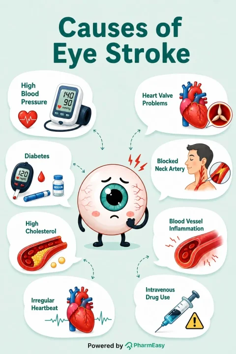
Stroke in eye may mainly occur due to disruption of the blood flow through the central retinal artery or vein due to its blockage by fat, calcium, or blood clot2,3,5. Eye stroke causes include2:
Eye stroke treatment includes various techniques to restore blood flow to the eye. However, treatment options are limited, and vision may not fully recover, especially when a large retinal area is involved. Time is critical, and treatment should be initiated as soon as possible. Studies suggest that treatment should be initiated within 2 to 4 hours of symptom onset2,5. Eye stroke treatment includes:
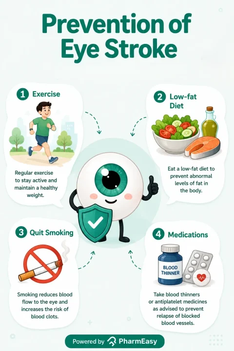
Measures to avoid an eye stroke include managing the conditions that affect the blood vessels, such as coronary artery disease2. This involves:
Also Read: Pain Behind Eye: Causes, Symptoms, Treatment, and Home Remedies
An eye stroke is a medical emergency, and you must seek prompt medical attention if you experience symptoms such as blurring or vision loss. If you are at risk of heart or blood vessel conditions, it is advised to routinely evaluate your vision as early detection can help in preserving your vision5.
Also Read: Could Yellow Eyes Be Serious? Causes, Symptoms & Treatment Guide
An eye stroke could happen suddenly without any warning. However, prompt intervention can play a significant role in treatment outcomes to protect the vision. Never ignore symptoms such as sudden vision loss, dark spots or blurring of vision, as they may require quick medical attention. Early diagnosis and adequate treatment could help to preserve the vision and detect any underlying medical condition that needs attention. If you or your dear ones experience any such eye stroke symptoms, seek swift emergency care, because when it comes to an eye stroke, every minute counts.
Also Read: What Is Eye Strain and How Can You Reduce It in the Digital Age
An eye stroke itself is not usually life-threatening. However, it may be a warning sign of underlying blood vessel disease and is associated with an increased risk of a brain stroke. Because both conditions share the same risk factors, prompt medical evaluation is essential6.
An eye stroke may cause permanent loss of vision, but the outcome depends on how severe the blockage is and how quickly treatment is started. Prompt treatment offers the best chance of preserving vision, although full recovery may not always be possible4,5.
Studies suggest a golden window for the treatment of eye stroke lies between 90 minutes and 4 hours from the beginning of symptoms however, full recovery may not always be possible8.
Stress alone may not directly cause an eye stroke; however, long-term stress may contribute to conditions such as high blood pressure and narrowing of blood vessels, which can increase the risk of an eye stroke9.
Eye stroke is quite rare, with data suggesting that around 1 in 10,000 people may experience it. It is also rarely reported in young individuals10.
An eye stroke does not directly lead to a brain stroke. However, it is associated with a higher risk of developing a brain stroke because both conditions share many of the same risk factors, such as high blood pressure and diabetes11.
Disclaimer: The information provided here is for educational/awareness purposes only and is not intended to be a substitute for medical treatment by a healthcare professional and should not be relied upon to diagnose or treat any medical condition. The reader should consult a registered medical practitioner to determine the appropriateness of the information and before consuming any medication. PharmEasy does not provide any guarantee or warranty (express or implied) regarding the accuracy, adequacy, completeness, legality, reliability or usefulness of the information; and disclaims any liability arising thereof.
Links and product recommendations in the information provided here are advertisements of third-party products available on the website. PharmEasy does not make any representation on the accuracy or suitability of such products/services. Advertisements do not influence the editorial decisions or content. The information in this blog is subject to change without notice. The authors and administrators reserve the right to modify, add, or remove content without notification. It is your responsibility to review this disclaimer regularly for any changes.
Many people, particularly young adults, want to gain muscle to feel stronger, improve fitness, or support healthy body goals1. However, building muscle takes more than exercise alone. Resistance training, along with a balanced muscle gain diet, gives the body the nutrients it needs to repair and build muscle after exercise. This repair process is often called muscle protein synthesis. Eating enough protein, carbohydrates, healthy fats, and other essential nutrients can support workout performance, muscle growth, and recovery2. This blog explains what a muscle gain diet is, the best foods to include, a simple 7-day meal plan, and practical nutrition tips to help you build muscle safely and effectively.
Muscles grow when the body repairs and builds new muscle tissue after exercise. Resistance training gives the muscles a reason to adapt, while nutrients from food provide the building blocks and energy needed for repair and growth. Protein intake and resistance exercise work together to support muscle development2.
Protein is important for muscle gain because it provides amino acids, which are the body’s building blocks for muscles. When you eat enough protein, your body is better able to repair muscle after exercise and builds new muscle tissue. Spreading protein across meals during the day may help provide a steady supply of amino acids for muscle recovery2. Diet also gives you the energy needed to exercise and recover. Carbohydrates help fuel workouts by supporting muscle glycogen, the stored form of carbohydrate used during exercise. After training, carbohydrates help refill these energy stores. Nutrient-rich foods, including milk and other good-quality protein sources, also provide vitamins and minerals that support muscle health3.
To build muscle, your body needs nutrients that help repair muscle, provide energy for training, and support recovery after workouts4,5.
Important nutrients for muscle gain include:
A varied diet that includes these nutrients can help your body respond well to resistance training and support long-term improvements in strength and muscle size4,5.
The best foods for muscle gain are foods that provide protein, energy, and other nutrients your body needs for training and recovery. Protein-rich foods are especially helpful because they provide amino acids for muscle repair and growth when combined with regular resistance exercise4,6.
Some of the best foods to gain muscle include:

Eggs are a good-quality protein source because they provide all the essential amino acids needed for muscle repair and growth2. They also contain leucine, an amino acid that helps support the muscle-building process2,4.

Milk contains whey and casein proteins, which provide essential amino acids for muscle repair and growth. Having milk after exercise may help support recovery and refill energy stores when it is part of a balanced diet2,6.
Pulses such as lentils, chickpeas, green gram, kidney beans, and peas provide plant-based protein. Combining pulses with cereals improves the balance of essential amino acids6.
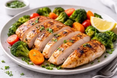
Fish, poultry, and lean meat provide complete proteins containing all essential amino acids required for muscle protein synthesis.
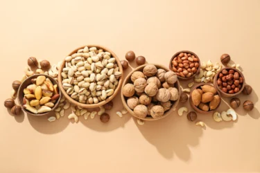
Nuts and seeds, including almonds, walnuts, chia seeds, pumpkin seeds, and flax seeds, provide plant-based protein and healthy fats.
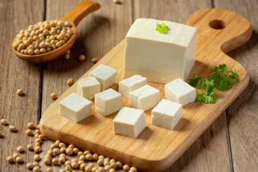
Soybeans and soy products provide plant-based protein containing essential amino acids4,6.
A muscle gain diet should focus on nutritious foods most of the time. It is helpful to limit foods that are high in added fat, sugar, and salt, as well as many ultra-processed foods, because they may add calories without providing many important nutrients.
Some foods to limit during a muscle gain diet include:
Adding vitamins or minerals to ultra-processed foods does not make them as nutritious as fresh, minimally processed foods6.
A balanced 7-day meal plan for muscle gain should include enough protein, carbohydrates, healthy fats, and a variety of nutrient-rich foods. These may include cereals, pulses, dairy products, eggs, lean meat or fish, fruits, vegetables, nuts, and seeds. Together, these foods can support muscle growth, recovery, and overall nutrition needs4,6.
The table below provides a sample 7-day meal plan for muscle gain that can be adjusted according to individual calorie needs, dietary preferences, and training goals2,4,6.
| Day | Breakfast | Lunch | Snacks | Dinner |
| Day 1 | Oats with milk, banana and nuts | Brown rice, dal, vegetables, curd and salad | Fruit smoothie with milk | Chapati, paneer/tofu, vegetables and curd |
| Day 2 | Millet porridge with milk, boiled eggs and fruit | Whole grain rice, chickpea curry, green leafy vegetables and curd | Nuts, fruit and yoghurt | Chapati, fish/chicken curry, vegetables and salad |
| Day 3 | Whole wheat bread, eggs and milk | Roti, rajma, vegetables, curd and fruit | Banana with peanut butter or nuts | Rice, dal, vegetables and curd |
| Day 4 | Ragi dosa with sambar and milk | Millet rice, pulses, vegetables and curd | Fruit smoothie with yoghurt | Chapati with paneer/chicken and vegetables |
| Day 5 | Oatmeal with milk, nuts and seasonal fruit | Rice, fish/egg curry, vegetables and salad | Sprouts, nuts and fruit | Whole grain roti, dal, vegetables and curd |
| Day 6 | Idli with sambar, milk and fruit | Chapati, soybean/pulse curry, vegetables and curd | Milk with banana or homemade smoothie | Rice, lean meat/fish or pulses, vegetables and salad |
| Day 7 | Millet upma with vegetables, nuts and milk | Whole grain rice, dal, vegetables, curd and fruit | Boiled eggs or roasted chickpeas with fruit | Chapati, paneer/tofu or chicken, vegetables and curd |
This meal plan focuses on obtaining essential nutrients from a variety of foods rather than relying on excessive supplements, helping support muscle growth, energy levels, and recovery during trainin2,5,6.
A gym diet plan for muscle gain should include the right foods before and after workouts to provide energy, support muscle repair, and improve recovery. The following sections explain what to eat before and after workouts:
| Meal | Sample foods |
| Breakfast | Oatmeal with a banana |
| Mid-morning snack | Low-fat yoghurt |
| Lunch | Whole-grain pasta |
| Pre-workout meal | Fruit smoothie made with low-fat milk |
| Post-workout meal | Whey protein shake with a banana |
| Dinner | Whole-grain cereal with milk |
| Bedtime snack | Fruits |
This is only a sample gym diet plan. You can change the foods and portion sizes to suit your daily routine, workout schedule, and personal food preferences5.
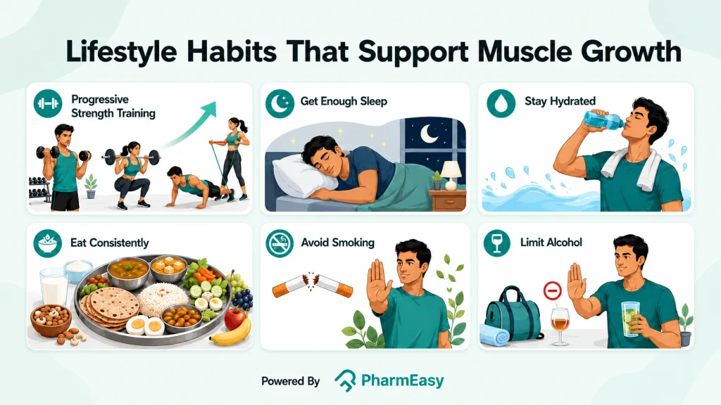
Building muscle is not only about diet. Healthy lifestyle habits also help your muscles recover, improve workout performance, and support steady progress over time. Important habits include:
Along with following a muscle gain diet, practising these healthy habits can help you build muscle more effectively and stay healthy.
Also Read: List of Protein Rich Food for Vegetarians
A well-planned muscle gain diet, combined with regular resistance training, can help support muscle growth, improve strength, and promote recovery. Focus on including high-quality protein, carbohydrates, healthy fats, and a variety of nutrient-rich whole foods instead of relying on supplements. Along with following a balanced diet, stay well hydrated, get enough sleep, manage stress, and avoid smoking and excessive alcohol consumption to support your muscle-building goals and overall health.
A balanced muscle gain diet that includes enough protein, carbohydrates, and healthy fats is best for supporting muscle growth and recovery. Protein helps repair and build muscle, while carbohydrates provide energy for training and help refill muscle energy stores5. For many active adults, about 1.4–2.0 g of protein per kilogram of body weight per day can support muscle growth when combined with resistance training2.
Muscle growth can be affected by smoking, heavy alcohol use, poor nutrition, not eating enough protein or calories, lack of regular strength training, and poor recovery14,15. Smoking is linked with lower muscle protein synthesis, while too much alcohol can slow muscle repair after exercise14.
Eating a protein-rich meal or snack every 3–4 hours can help support muscle protein synthesis throughout the day. After resistance exercise, eating high-quality protein soon after your workout may help muscles recover and grow over time2.
Two meals a day may make it harder for many people to meet their protein and energy needs for muscle gain. Spreading protein across several meals or snacks during the day may better support muscle repair and growth16.
Good snacks for muscle growth include low-fat yoghurt, fruit smoothies, bananas with a whey protein shake, and whole-grain cereal with milk. These snacks provide protein and carbohydrates, which help support muscle recovery and replenish energy after exercise5.
1. Ganson KT, Nguyen L, Ali ARH, et al. “Eat more protein, build more muscle”: A grounded theory study of muscle-building behaviors among Canadian adolescents and young adults. Body Image. 2023;47:101635. doi:10.1016/j.bodyim.2023.101635. Available from: https://pubmed.ncbi.nlm.nih.gov/37806066/
2. Jäger R, Kerksick CM, Campbell BI, et al. International Society of Sports Nutrition Position Stand: protein and exercise. J Int Soc Sports Nutr. 2017;14(1):20. doi:10.1186/s12970-017-0177-8. Available from: https://link.springer.com/article/10.1186/s12970-017-0177-8
3. Moore DR, Philp A. Editorial: Nutritional Strategies to Promote Muscle Mass and Function Across the Health Span. Front Nutr. 2020;7:569270. doi:10.3389/fnut.2020.569270. Available from: https://pmc.ncbi.nlm.nih.gov/articles/PMC7561707/
4. Iraki J, Fitschen P, Espinar S, Helms E. Nutrition Recommendations for Bodybuilders in the Off-Season: A Narrative Review. Sports. 2019;7(7):154. doi:10.3390/sports7070154. Available from: https://www.researchgate.net/publication/334044740_Nutrition_Recommendations_for_Bodybuilders_in_the_Off-Season_A_Narrative_Review
5. Hwang DJ, Yang HJ. Nutritional Strategies for Enhancing Performance and Training Adaptation in Weightlifters. Int J Mol Sci. 2024;26(1):240. doi:10.3390/ijms26010240. Available from: https://www.researchgate.net/publication/387572639_Nutritional_Strategies_for_Enhancing_Performance_and_Training_Adaptation_in_Weightlifters
6. ICMR–National Institute of Nutrition. Dietary Guidelines for Indians – 2024. Indian Council of Medical Research (ICMR), Department of Health Research, Ministry of Health and Family Welfare, Government of India; 2024. Accessed July 24, 2026. Available from: https://nin.res.in/dietaryguidelines/pdfjs/locale/DGI_2024.pdf
7. Strength training for beginners | healthdirect. Accessed July 24, 2026. Available from: https://www.healthdirect.gov.au/strength-training-for-beginners
8. Knowles OE, Drinkwater EJ, Urwin CS, Lamon S, Aisbett B. Inadequate sleep and muscle strength: Implications for resistance training. J Sci Med Sport. 2018;21(9):959-968. doi:10.1016/j.jsams.2018.01.012. Available from: https://europepmc.org/article/med/29422383
9. Exercise and Fluid Replacement. Med Sci Sports Exerc. 2007;39(2):377-390. doi:10.1249/mss.0b013e31802ca597. Available from: https://www.ovid.com/jnls/acsm-msse/abstract/10.1249/mss.0b013e31802ca597~exercise-and-fluid-replacement?redirectionsource=fulltextview
10. Helms ER, Aragon AA, Fitschen PJ. Evidence-based recommendations for natural bodybuilding contest preparation: nutrition and supplementation. J Int Soc Sports Nutr. 2014;11(1):20. doi:10.1186/1550-2783-11-20. Available from: https://pmc.ncbi.nlm.nih.gov/articles/PMC4033492/
11. Rom O, Kaisari S, Aizenbud D, Reznick AZ. Lifestyle and Sarcopenia – Etiology, Prevention and Treatment. Rambam Maimonides Med J. 2012;3(4):e0024. doi:10.5041/RMMJ.10091. Available from: https://pmc.ncbi.nlm.nih.gov/articles/PMC3678825/
12. Centers for Disease Control and Prevention. How to Quit Smoking. May 2024. Available from: https://www.cdc.gov/tobacco/media/pdfs/2024/05/cdc-osh-how-to-quit-smoking-infographic-web-508.pdf
13. CDC. What You Can Do to Lower Your Risk of Alcohol-Related Harms. Alcohol Use. February 25, 2025. Accessed July 24, 2026. Available from: https://www.cdc.gov/alcohol/about-alcohol-use/lower-alcohol-related-harms.html
14. Parr EB, Camera DM, Areta JL, et al. Alcohol Ingestion Impairs Maximal Post-Exercise Rates of Myofibrillar Protein Synthesis following a Single Bout of Concurrent Training. Alway SE, ed. PLoS ONE. 2014;9(2):e88384. doi:10.1371/journal.pone.0088384. Available from: https://journals.plos.org/plosone/article?id=10.1371/journal.pone.0088384
15. Petersen AMW, Magkos F, Atherton P, et al. Smoking impairs muscle protein synthesis and increases the expression of myostatin and MAFbx in muscle. Am J Physiol-Endocrinol Metab. 2007;293(3):E843-E848. doi:10.1152/ajpendo.00301.2007. Available from: https://www.researchgate.net/publication/6229004_Smoking_impairs_muscle_protein_synthesis_and_increases_the_expression_of_myostatin_and_MAFbx_in_muscle
16. Schoenfeld BJ, Aragon AA. How much protein can the body use in a single meal for muscle-building? Implications for daily protein distribution. J Int Soc Sports Nutr. 2018;15(1):10. doi:10.1186/s12970-018-0215-1. Available from: https://pmc.ncbi.nlm.nih.gov/articles/PMC5828430/
Disclaimer: The information provided here is for educational/awareness purposes only and is not intended to be a substitute for medical treatment by a healthcare professional and should not be relied upon to diagnose or treat any medical condition. The reader should consult a registered medical practitioner to determine the appropriateness of the information and before consuming any medication. PharmEasy does not provide any guarantee or warranty (express or implied) regarding the accuracy, adequacy, completeness, legality, reliability or usefulness of the information; and disclaims any liability arising thereof.
Links and product recommendations in the information provided here are advertisements of third-party products available on the website. PharmEasy does not make any representation on the accuracy or suitability of such products/services. Advertisements do not influence the editorial decisions or content. The information in this blog is subject to change without notice. The authors and administrators reserve the right to modify, add, or remove content without notification. It is your responsibility to review this disclaimer regularly for any changes.