You’ve probably heard of this disclaimer on multiple occasions – ‘smoking is injurious to health’. What you may not immediately realise is the extent of the damage smoking can cause to your health and, most directly, your lungs.
Smoking is known to be the leading cause of preventable diseases and deaths globally. Nearly all forms of lung cancer, the top cause of cancer death in both men and women, can be attributed to smoking. Tobacco and tobacco-related products can damage the lungs’ ability to supply oxygen to the body. Other substances commonly found in cigarette smoke can cause permanent lung damage, even in small amounts.
A single puff of cigarette smoke contains upwards of 7,000 chemicals. Tobacco smoke contains over 70 known cancer-causing chemicals2. When you breathe these in, these toxins go deep into your lungs and can cause swelling, resulting in a host of other respiratory diseases.
Both tobacco and chemical substances found in cigarettes can change the cellular structure of the lungs. They can cause the elastic walls within the airways to break down – resulting in less functioning surface area in the lungs. Cigarettes can damage lung tissue, preventing them from functioning correctly. This can increase the risk of diseases caused by smoking, such as chronic bronchitis, emphysema, respiratory diseases, asthma and COPD (Chronic Obstructive Pulmonary Disease)1.
Nicotine in tobacco can also damage the ability of the respiratory system to filter out dust and dirt. This can lead to toxic substances passing through, resulting in lung congestion and the ‘smoker’s cough’.
Also Read: What Is Hantavirus? Symptoms, Causes, & Effective Prevention
A person who smokes throughout life is at high risk of developing a range of potentially fatal diseases owing to impaired lung function and breathlessness due to swelling and narrowing of the lung airways and excess mucus build-up. They are also prone to weakening the lungs’ clearance system, leading to the accumulation of toxic substances and causing lung irritation and damage. Further, they are also at an increased risk of lung infection, chronic bronchitis and heightened risk of asthma, along with permanent damage to air sacs3.
In the longer term, smoking is known to induce heart disease and stroke, in certain cases, it can cause ulcers of the digestive system and put smokers at increased risk of type 2 diabetes.
Most smokers are also likely to develop emphysema. The number of cigarettes you smoke and other lifestyle factors may impact the extent of the damage. If you’re diagnosed with either of these respiratory diseases – emphysema or chronic bronchitis, you run the risk of being diagnosed with chronic obstructive pulmonary disease (COPD).
Also Read: Does Smoking Really Affect Your Brain?
Smoking can affect a person’s health in other ways, too, harming almost every organ in the body. In most cases, it can result in a compromised immune system function, making you susceptible to many other illnesses. It can also lead to lower bone density (brittle bones), which increases the risk of broken bones and fractures. Smoking also leaves you at a higher risk of rheumatoid arthritis, heart disease and stroke, along with an increased risk for cataracts (clouding of the eye lenses).
Apart from respiratory diseases, other visible disorders include an increased risk of oral cancers, gum disease and tooth loss, premature ageing of the skin, bad breath and stained teeth and an increased risk for age-related macular degeneration, which can lead to blindness. Moreover, even your wounds may take longer to heal!
Also Read: 6 Simple Exercises to Improve Your Lung Health
It’s never too late to quit smoking. Within days of quitting smoking, lungs begin to repair themselves. In fact, just 12 hours after you quit, the amount of carbon monoxide in your blood drops to a much healthier level. More oxygen flows to your vital organs and you will be able to breathe better. In about 10 to 15 years, your risk of developing lung cancer reduces and may even become the same as a non-smoker4.
Also Read: How to Avoid Asthma Attacks During Winter
1. Centers for Disease Control and Prevention (US); National Center for Chronic Disease Prevention and Health Promotion (US); Office on Smoking and Health (US). How Tobacco Smoke Causes Disease: The Biology and Behavioral Basis for Smoking-Attributable Disease: A Report of the Surgeon General. Atlanta (GA): Centers for Disease Control and Prevention (US); 2010. 7, Pulmonary Diseases. Available from: https://www.ncbi.nlm.nih.gov/books/NBK53021/
2. National Cancer Institute. Harms of Cigarette Smoking and Health Benefits of Quitting [Internet]. Bethesda (MD): National Cancer Institute; reviewed 19 December 2017 [cited 2025 Sep 19]. Available from: https://www.cancer.gov/about-cancer/causes-prevention/risk/tobacco/cessation-fact-sheet
3. Varghese J, Muntode Gharde P. A Comprehensive Review on the Impacts of Smoking on the Health of an Individual. Cureus. 2023 Oct 5;15(10):e46532. doi: 10.7759/cureus.46532. PMID: 37927763; PMCID: PMC10625450. Available from: https://pmc.ncbi.nlm.nih.gov/articles/PMC10625450/
4. Centers for Disease Control and Prevention. Benefits of Quitting Smoking [Internet]. Atlanta (GA): CDC; updated May 15, 2024 [cited 2025 Sep 19]. Available from: https://www.cdc.gov/tobacco/about/benefits-of-quitting.html
Disclaimer: The information provided here is for educational/awareness purposes only and is not intended to be a substitute for medical treatment by a healthcare professional and should not be relied upon to diagnose or treat any medical condition. The reader should consult a registered medical practitioner to determine the appropriateness of the information and before consuming any medication. PharmEasy does not provide any guarantee or warranty (express or implied) regarding the accuracy, adequacy, completeness, legality, reliability or usefulness of the information; and disclaims any liability arising thereof.
Links and product recommendations in the information provided here are advertisements of third-party products available on the website. PharmEasy does not make any representation on the accuracy or suitability of such products/services. Advertisements do not influence the editorial decisions or content. The information in this blog is subject to change without notice. The authors and administrators reserve the right to modify, add, or remove content without notification. It is your responsibility to review this disclaimer regularly for any changes.
 10
10

31st May is known as the ”World No Tobacco Day” and for a good reason too1. Did you know that more than 10 million die each year in India due to tobacco? India is home to 12% of the world’s smokers, according to the World Health Organization (WHO). You have heard numerous people tell you that smoking affects your lungs. You have seen the gross pictures on cigarette packs but smoked anyway. But did you know that smoking affects your brain too?
Nicotine works like the various neurotransmitters that are already there in our brain. It activates dopamine signals that result in a pleasant sensation in your brain. With the passing of time and more smoking, the brain reduces acetylcholine receptors to compensate for the increased signalling activity. As a result, nicotine tolerance is created in the brain2.
The brain ends up needing more nicotine. As nicotine mimics the work of dopamine that provides the feel-good factor, your brain starts associating smoking (nicotine use) with feeling good. The nicotine in cigarettes changes your brain and makes you suffer from withdrawal symptoms when you try to quit. You start feeling irritable, anxious, and your body has a strong craving for nicotine. As a result of these symptoms, most people reach for another cigarette, and then another and are unable to quit.

Brain size and volume is associated with higher intelligence and better cognitive functioning. The average brain volume in adult males is 1260 cubic cm and 1130 cubic cm in adult females. According to a 2017 study2, the longer you smoke, the more your brain loses volume with vital tissues shrivelling up.
Smoking affects the subcortical brain regions. The subcortical areas of the brain are associated with pleasure, hormone production, emotion, and memory. Smokers thus develop age-related loss of brain volume that leads to an increased risk of dementia and is one of the ways how smoking harms the brain.

Dementia is a syndrome that is characterized by deterioration in thinking, memory, behaviour, and the ability to perform everyday activities. It is said to affect older people mainly, but it is not a normal part of ageing. Since smoking affects the subcortical regions of the brain that are associated with memory, it puts smokers at a higher risk of dementia.
In 2015, a research team reviewed 37 studies that compared smokers and non-smokers and found that smokers were 30 % more likely to be affected by dementia. Quitting smoking can decrease the risk of dementia in the person4.
Cigarette smoking has been associated with dementia and dementia-related brain changes, notably gray matter (GM) volume atrophy. These associations are thought to reflect the co-morbidity of smoking and vascular, respiratory, and substance use/psychological conditions.
Dr. M.G. Kartheeka, MBBS, MD(Pediatrics)

One of the smoking effects on brain is cognitive decline, which usually happens as people get older. But in smokers, it starts much earlier. Signs and symptoms of cognitive decline include:
In 2012, the cognitive data of about 7,000 men and women were studied for 12 years. The researchers found that smokers experienced a much more rapid cognitive decline than non-smokers. Middle-aged male smokers were found to be more at risk than female smokers4.
If you smoke say, 20 cigarettes a day, you are 6 times more likely to have a stroke than a non-smoker. Tobacco contains over 7,000 harmful chemicals, including formaldehyde, cyanide, arsenic, and carbon monoxide. These toxic chemicals get transferred from the lungs to the blood. They make platelets more likely to stick together. Platelets help in clotting the blood in case of blood loss, but if the platelets stick together, it increases the chance of clot-forming5.
Smokers are at a higher risk of developing atherosclerosis where arteries become hardened and narrow. It restricts smooth blood flow making the formation of blood clots more likely. If a clot forms in an artery leading to the brain, it can block the blood supply to a part of the brain resulting in a stroke. This is known as ischaemic stroke. Smoking is said to double the risk of having an ischaemic stroke. If a person quits smoking, within 5 years, his/her risk of stroke will start decreasing to that of a non-smoker.

Smoking releases a severe amount of toxicity in our bodies. There are about 60 known cancer-causing substances in tobacco6. The chemicals that make up a cigarette are:
Smoking also causes a temporary spike in blood pressure, which can weaken the arterial walls and make them more prone to form an aneurysm and rupture. The harmful chemicals in a cigarette are also implicated in the causation of brain cancer.
Dr. Ashish Bajaj, M.B.B.S., M.D. in Clinical Pharmacology and Toxicology

Smoking affects the brain and hence, mental health. Sometimes, bad mental health makes people take up smoking and worsen their conditions. Other times, it is the other way around7.

The nicotine from cigarettes alters the brain. It makes the brain connect ‘feeling good’ to smoking. Quitting smoking becomes tough after some time because smokers start suffering from withdrawal symptoms. They then find solace in smoking and fall prey to the dangerous cycle and become addicted.

How many times have you heard somebody say, ‘I’m feeling stressed out, I need to smoke right now’ or ‘Smoking makes me feel relaxed’7?
Stress is very common and can cause symptoms like headaches, irritability, anxiety, and/or breathlessness at times. Smoking increases the occurrence of these symptoms. Smokers start feeling the symptoms if they do not smoke for a long time and associate smoking with being a reliever of stress.

Nicotine mimics the work of dopamine, prompting the brain to switch off its mechanism that makes and secretes dopamine. In the long term, the supply of dopamine decreases in the brain and inspires people to smoke more. There is a complex relationship between depression and smoking. Smokers with depression have more trouble quitting as withdrawal symptoms become more severe in them7.

Research has shown smoking increases tension and anxiety. The relaxed feeling that smokers talk about after a quick smoke fades away just as quickly. It is hugely short-lived and only adds more jitteriness in the smoker, making him/her reach for more7.

It has been reported that people who suffer from a serious mental disorder known as Schizophrenia tend to be heavy smokers. Some people suffering from this disorder have claimed that smoking helps them to numb the debilitating symptoms of schizophrenia and also to mitigate the side effects experienced from the medication for the same. Ironically, recent research has found that excessive smoking may very well be one of the causes for the onset of schizophrenia. However, since there is more research required to fully confirm this, it has not yet received mainstream acceptance. Nevertheless, it is best to avoid smoking to reduce the risk of developing such mental disorders8.
Yes, e-cigarettes have negative effects on the brain too. National Institute on Drug Abuse has reported that the nicotine in e-cigarettes goes about making similar harmful changes in the brain. E-cigarette vapour contains harmful chemicals too hence it is not a way out.
If all this information on how smoking affects the brain has you worried, you can always try quitting. Most addictions are hard to overcome. But since smoking has been around for a while there are well-established methods to try out. Keep in mind, since everyone is different not all approaches will work the same for you. Some may be more effective than others, do what works best for you9.
Absolutely! Within 20 minutes of quitting smoking, your heart rate will slow down. Within 12 hours, levels of carbon monoxide in your blood will start decreasing. Within 3 months, lung functions and blood circulation will start getting better. Within a year of quitting, your risk of having a heart attack will start decreasing by a whopping 50 %. Within 5 to 15 years, your risk of suffering a stroke will reduce to that of a non-smoker.
Also Read: What Happens To Your Lungs From Smoking? Things You Should Know
1. World Health Organization. World No Tobacco Day – 31 May is World No Tobacco Day [Internet]. Geneva: WHO; [cited 2025 Dec 5]. Available from: https://www.who.int/campaigns/world-no-tobacco-day
2. Valentine G, Sofuoglu M. Cognitive Effects of Nicotine: Recent Progress. Curr Neuropharmacol. 2018;16(4):403-414. doi: 10.2174/1570159X15666171103152136. PMID: 29110618; PMCID: PMC6018192. Available from: https://pmc.ncbi.nlm.nih.gov/articles/PMC6018192/
3. Chang Y, Thornton V, Chaloemtoem A, Anokhin AP, Bijsterbosch J, Bogdan R, Hancock DB, Johnson EO, Bierut LJ. Investigating the Relationship Between Smoking Behavior and Global Brain Volume. Biol Psychiatry Glob Open Sci. 2023 Oct 6;4(1):74-82. doi: 10.1016/j.bpsgos.2023.09.006. PMID: 38130847; PMCID: PMC10733671. Available from: https://pmc.ncbi.nlm.nih.gov/articles/PMC10733671/
4. Peters R, Poulter R, Warner J, Beckett N, Burch L, Bulpitt C. Smoking, dementia and cognitive decline in the elderly, a systematic review. BMC Geriatr. 2008 Dec 23;8:36. doi: 10.1186/1471-2318-8-36. PMID: 19105840; PMCID: PMC2642819. Available from:https://pmc.ncbi.nlm.nih.gov/articles/PMC2642819/
5. Shah RS, Cole JW. Smoking and stroke: the more you smoke the more you stroke. Expert Rev Cardiovasc Ther. 2010 Jul;8(7):917-32. doi: 10.1586/erc.10.56. PMID: 20602553; PMCID: PMC2928253. Available from: https://pmc.ncbi.nlm.nih.gov/articles/PMC2928253/
6. Vida S, Richardson L, Cardis E, Krewski D, McBride M, Parent ME, Abrahamowicz M, Leffondré K, Siemiatycki J. Brain tumours and cigarette smoking: analysis of the INTERPHONE Canada case-control study. Environ Health. 2014 Jun 27;13:55. doi: 10.1186/1476-069X-13-55. PMID: 24972852; PMCID: PMC4088305. Available from: https://pmc.ncbi.nlm.nih.gov/articles/PMC4088305/
7. Boksa P. Smoking, psychiatric illness and the brain. J Psychiatry Neurosci. 2017 May;42(3):147-149. doi: 10.1503/jpn.170060. PMID: 28440208; PMCID: PMC5403659. Available from: https://pmc.ncbi.nlm.nih.gov/articles/PMC5403659/
8. Ding JB, Hu K. Cigarette Smoking and Schizophrenia: Etiology, Clinical, Pharmacological, and Treatment Implications. Schizophr Res Treatment. 2021 Dec 13;2021:7698030. doi: 10.1155/2021/7698030. PMID: 34938579; PMCID: PMC8687814. Available from: https://pmc.ncbi.nlm.nih.gov/articles/PMC8687814/
9. Centers for Disease Control and Prevention. Tips For Quitting. Tips From Former Smokers. 2024 Sept 27 [cited 2025 Dec 05]. Available from: https://www.cdc.gov/tobacco/campaign/tips/quit-smoking/tips-for-quitting/index.html
Disclaimer: The information provided here is for educational/awareness purposes only and is not intended to be a substitute for medical treatment by a healthcare professional and should not be relied upon to diagnose or treat any medical condition. The reader should consult a registered medical practitioner to determine the appropriateness of the information and before consuming any medication. PharmEasy does not provide any guarantee or warranty (express or implied) regarding the accuracy, adequacy, completeness, legality, reliability or usefulness of the information; and disclaims any liability arising thereof.
Links and product recommendations in the information provided here are advertisements of third-party products available on the website. PharmEasy does not make any representation on the accuracy or suitability of such products/services. Advertisements do not influence the editorial decisions or content. The information in this blog is subject to change without notice. The authors and administrators reserve the right to modify, add, or remove content without notification. It is your responsibility to review this disclaimer regularly for any changes.
20
2
Headaches are among the most common health conditions, affecting individuals of all ages across the world. According to the World Health Organisation (WHO), headache disorders affect over 40% of the world’s population1. While many people have occasional headaches, regular episodes can interfere with work, sleep, and other everyday activities. Understanding the different types of headaches can help identify their causes and improve their treatment and prevention2. In this blog, we will discuss how many types of headaches are there, their causes, how they are diagnosed, their treatment options, and simple tips to prevent them.
A headache is a pain or discomfort felt in the head, scalp, or face. It can range from mild to severe and may last for a few minutes or several hours2. Headaches occur when pain-sensitive structures in and around the head, such as blood vessels, nerves, muscles, and tissues, become irritated or triggered due to factors such as smell, stress, and medications2,3.
Headaches, especially when they occur frequently, can affect a person’s quality of life and even cause problems at work. The fear of getting another headache can greatly affect family relationships, social activities, and job performance. Living with long-term headaches may also increase the risk of other health problems, such as anxiety and depression, which is more commonly observed in people with migraines, a type of headache1.
There are two types of headaches: primary headaches and secondary headaches. The table below highlights the key differences between these two types:
| Feature | Primary Headaches | Secondary Headaches |
| Definition | Headaches that are not caused by any medical condition, but are a part of a headache disorder4. | Headaches that occur due to an underlying health problem5. |
| Common causes | Occurs due to complex changes in the brain and nervous system mechanisms that are involved in sensing and processing pain4. Factors such as stress, hormonal changes, lack of sleep, certain foods, and genetics can act as a trigger that brings on headache episodes2. | Occurs when an underlying health condition irritates or puts pressure on the pain-sensitive structures in the head and neck (such as nerves, brain covering [meninges], blood vessels, muscles and tissues covering the skull). These conditions can include brain infections, head injuries, medication overuse, extremely high blood pressure, acute sinus infections (long-term infections rarely cause headaches), strokes, tumours, and other brain disorders2,5. |
| Examples | Migraine, tension headache, cluster headache, etc.4. | Sinus headache, medication-overuse headache, cervicogenic headache, etc5. |
| Severity | Usually not life-threatening, but can interfere with quality of life1,4. | Depends on the cause and can range from mild to life-threatening5. |
| Treatment | Symptom management, trigger avoidance and prescription medications if needed2. | Treating underlying health condition5. |
Understanding the different types of headaches and their causes can help you identify possible triggers, recognise warning signs, and seek the right treatment when needed. Here are some of the common types of headaches:

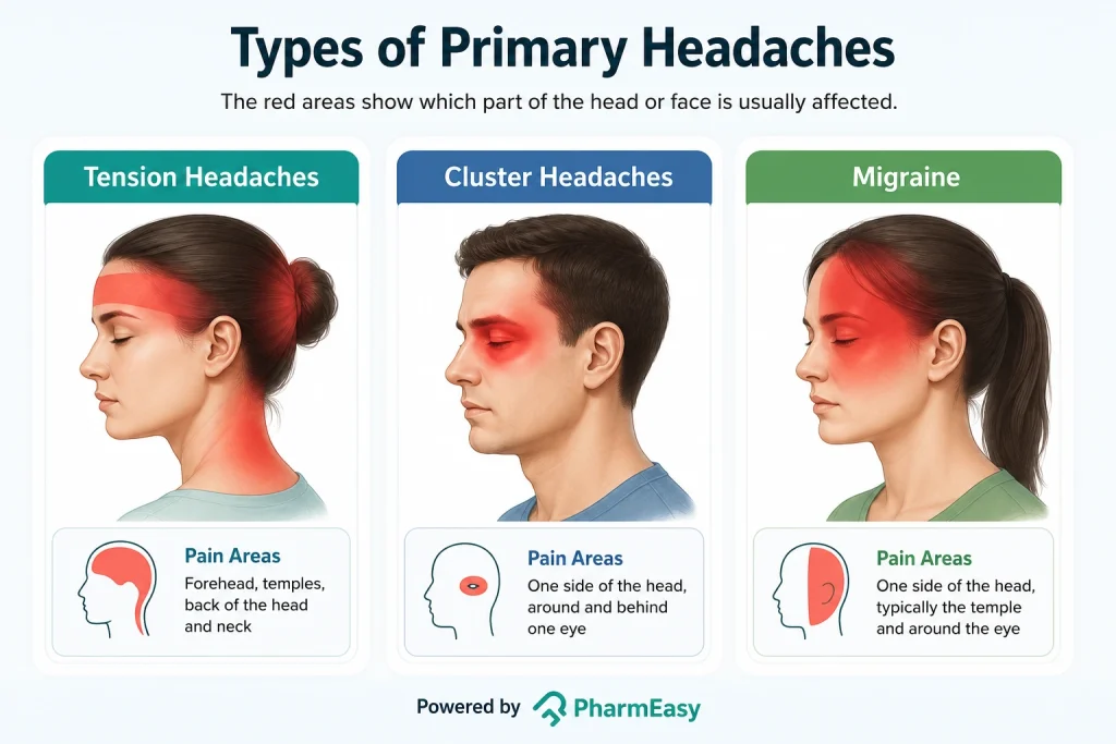
These are among the most common types of primary headaches. They usually last between 30 minutes and one week. Although they can start around puberty, they are most common in people in their 30s. Women and young people are more likely to experience this headache.

This is a type of primary headache that occurs repeatedly2. It can last between 4 and 72 hours. It often begins during puberty and is most common in people aged 35 to 45 years. Women are more likely to be affected than men due to hormonal issues. Migraine attacks in children are usually shorter than in adults1.
Note: Food triggers can vary from person to person. Alcohol (especially red wine), excess caffeine intake or suddenly stopping caffeine, chocolate, fish, and processed meats may trigger migraines in some people.
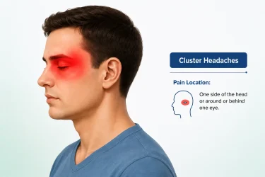
These are extremely painful but rare types of primary headaches. They can last from 15 minutes to 3 hours. The pain occurs in clusters or as repeated episodes, at the same time each day or night for many weeks. They most commonly occur between the ages of 20 and 40 years but can occur at any age, including in children and older adults2. These are more common in men than in women7.
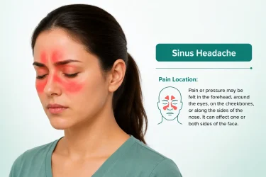
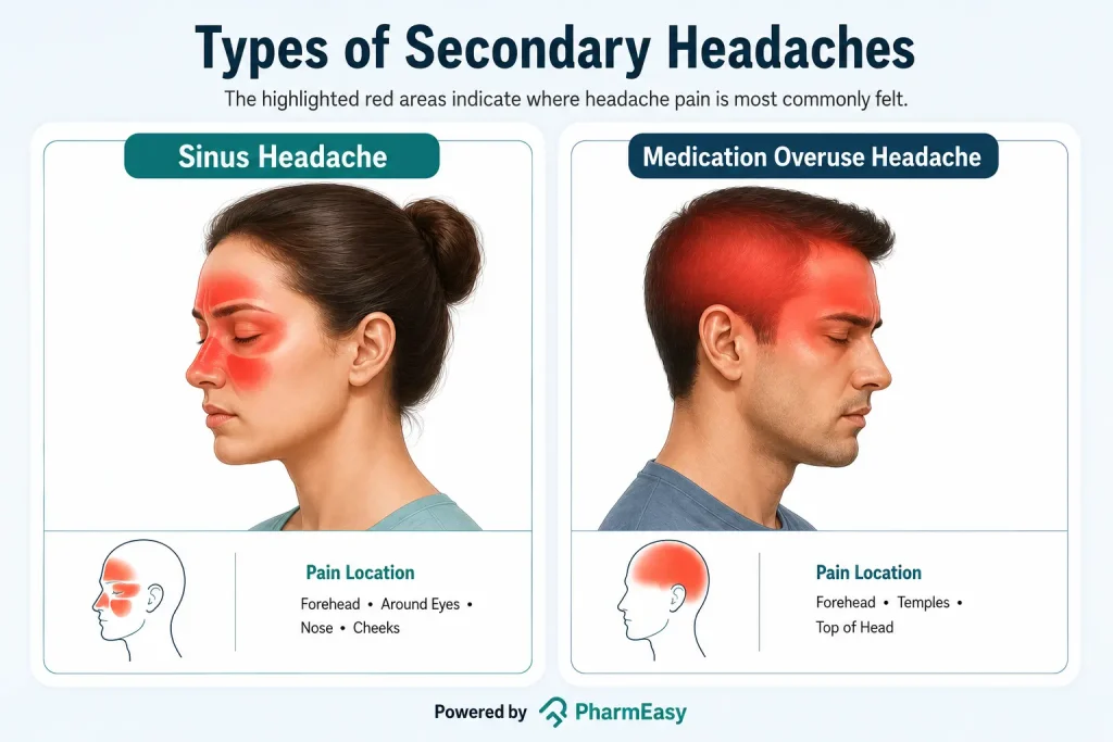
This is a type of secondary headache caused by the sinus issues9. Without treatment, the pain can persist for 4 weeks or less than that and become worse when a person bends forward or lies down. However, in most cases, the headache usually improves as the infection subsides9,10.
Note: Many headaches that people believe are caused by sinus problems are actually migraines. This is a common misunderstanding about headaches7.
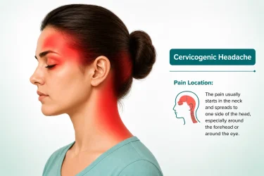
This is a type of secondary headache, which is rare and chronic. It is most commonly seen in people aged between 30 and 44 years. It is often mistaken for primary headaches, such as migraine and tension-type headache. The pain goes away when the underlying condition is treated successfully.
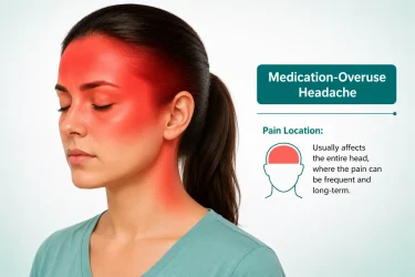
This is a type of secondary headache caused by the overuse of medications. It is also known as rebound headache. Medication-overuse headache is characterised as:
As there are different types of headaches and their reasons vary, doctors might use more than one diagnostic procedure to determine the cause of a headache. These include:
Note: High BP causes headache only when it causes hypertensive emergencies or BP rises suddenly. Mild or moderate high blood pressure does not usually cause headaches.
Note: If the doctor suspects that the increased pressure inside the skull is caused by a tumour or another growth, they may recommend a CT scan or MRI before considering a lumbar puncture. In some cases, a lumbar puncture may not be safe.
The treatment options usually available for headaches include:
Frequent headaches can often be managed by identifying and avoiding the triggers that cause them. The following measures may help lower the risk of recurrent headaches:


Also Read: Home Remedies For Headache By Dr. Siddharth Gupta
It is very important to consult a doctor if the headache:
Headaches are a common ailment and their severity can range from a mild ache to severe pain that interferes with daily activities. While most headaches are harmless, some may be a sign of an underlying health problem that needs medical attention. Knowing the different types of headaches, their symptoms, causes, and treatments can help you manage them better and recognise when to see a doctor.
The 5 C’s of headaches are common triggers that can worsen or cause headaches: Cheese, chocolate, citrus fruits, caffeine, and coke. Avoiding these triggers may help reduce headaches in some people, especially those with migraines16.
The type of headache can often be identified by its location, how the pain feels, how long it lasts, and its symptoms (such as nausea, sensitivity to light, or nasal congestion). A doctor might correlate these along with the individual’s medical history and triggers and, in some cases, recommend tests to rule out serious underlying conditions2,10.
Headaches that start suddenly and are severe; occur after a head injury; or are accompanied by symptoms such as weakness, confusion, seizures, fever, vision changes, or difficulty speaking require immediate medical attention as they may be a sign of serious conditions such as a stroke, meningitis, or bleeding in the brain2,5.
Consult a doctor immediately if your headaches are sudden or severe, occur after a head injury, or are associated with fever, confusion, weakness, vision problems, seizures, or difficulty speaking. These symptoms may be a sign of a serious condition and should not be ignored2.
A brain tumour-related headache is usually persistent, gradually worsens over time, and may be worse in the morning or with coughing and straining. It is important to note that these symptoms may also occur if a person has a migraine or cluster headache. A person with tumour may have other associated symptoms, such as seizures, vision changes, weakness, or difficulty speaking. Most headaches are not caused by brain tumours; however, a new or worsening headache with neurological symptoms should be evaluated by a doctor5.
1. Migraine and other headache disorders. 2025. Available from: https://www.who.int/news-room/fact-sheets/detail/headache-disorders
2. Headache. 2026. Available from: https://www.ninds.nih.gov/health-information/disorders/headache
3. The anatomy of head pain. In: Handbook of Clinical Neurology. Vol 198. Elsevier; 2023:41-60. doi:10.1016/B978-0-12-823356-6.00001-9 Available from: https://pubmed.ncbi.nlm.nih.gov/38043970/
4. Wang Z, Yang X, Zhao B, Li W. Primary headache disorders: From pathophysiology to neurostimulation therapies. Heliyon. 2023;9(4):e14786. doi:10.1016/j.heliyon.2023.e14786 Available from: https://pubmed.ncbi.nlm.nih.gov/37077680/
5. Wijeratne T, Wijeratne C, Korajkic N, Bird S, Sales C, Riederer F. Secondary headaches – red and green flags and their significance for diagnostics. eNeurologicalSci. 2023;32:100473. doi:10.1016/j.ensci.2023. 100473 Available from: https://pmc.ncbi.nlm.nih.gov/articles/PMC10339125/
6. Nihir Shah, Asuncion RMD, Hameed S. Muscle Contraction Tension Headache. 2024. Available from: https://www.ncbi.nlm.nih.gov/books/NBK562274/
7. Headache: When to worry, what to do. 2024. Available from: https://www.health.harvard.edu/pain/headache-when-to-worry-what-to-do
8. Cluster headache. 2025. Available from: https://medlineplus.gov/ency/article/000786.htm
9. Sinusitis. 2024. Available from: https://medlineplus.gov/ency/article/000647.htm
10. Headaches. 2025. Available from: https://www.healthdirect.gov.au/headaches
11. Al Khalili Y, K. Ly N, Murphy PB. Cervicogenic Headache. 2022. Available from: https://www.ncbi.nlm.nih.gov/books/NBK507862/
12. Aleksenko D, Lui F, Sánchez-Manso JC. Medication Overuse Headache. 2025. Available from: https://www.ncbi.nlm.nih.gov/sites/books/NBK470171/
13. Medication Overuse Headache. 2022. Available from: https://headaches.org/resources/medication-overuse-headache/
14. Kim KT. Lumbar puncture: considerations, procedure, and complications. encephalitis. 2022;2(4):93-97. doi:10.47936/encephalitis.2022.00045 Available from: https://pmc.ncbi.nlm.nih.gov/articles/PMC10295920/
15. Eskey CJ, Ogilvy CS. Fluoroscopy-guided lumbar puncture: decreased frequency of traumatic tap and implications for the assessment of CT-negative acute subarachnoid hemorrhage. AJNR Am J Neuroradiol. 2001;22(3):571-576. Available from: https://pubmed.ncbi.nlm.nih.gov/11237986/
16. Speight N. Best practice: migraine. Arch Dis Child. 2006 Jun;91(6):541. PMID: 16714736; PMCID: PMC2082764. Available from: https://pmc.ncbi.nlm.nih.gov/articles/PMC2082764/
Disclaimer: The information provided here is for educational/awareness purposes only and is not intended to be a substitute for medical treatment by a healthcare professional and should not be relied upon to diagnose or treat any medical condition. The reader should consult a registered medical practitioner to determine the appropriateness of the information and before consuming any medication. PharmEasy does not provide any guarantee or warranty (express or implied) regarding the accuracy, adequacy, completeness, legality, reliability or usefulness of the information; and disclaims any liability arising thereof.
Links and product recommendations in the information provided here are advertisements of third-party products available on the website. PharmEasy does not make any representation on the accuracy or suitability of such products/services. Advertisements do not influence the editorial decisions or content. The information in this blog is subject to change without notice. The authors and administrators reserve the right to modify, add, or remove content without notification. It is your responsibility to review this disclaimer regularly for any changes.

Kidney disease often develops quietly, with little or no warning in its early stages. That is why many individuals do not realise there is a problem until the condition has progressed to an advanced stage1.
However, recognising the early signs of kidney disease and seeking timely medical care can help slow or even avoid further kidney damage.
In this blog, we will explore the early kidney disease symptoms, types and causes, lifestyle and prevention tips, and when it is important to consult a doctor.
Healthy kidneys filter waste products, excess fluid, and toxins from the blood. They also help regulate blood pressure, maintain the body’s fluid and mineral balance, and produce hormones that support bone health and red blood cell production2. Kidney disease occurs when the structure or function of the kidney is impaired. This can cause waste and fluid to build up in the body3,4.
Did You Know?
Depending on the rate at which the symptoms have developed, kidney disease can be of two main types4:
The seriousness of your kidney disease is primarily determined by your eGFR (estimated glomerular filtration rate), which measures how effectively your kidneys filter waste from your blood, and your uACR (urine albumin-to-creatinine ratio), which assesses kidney damage and protein leakage.
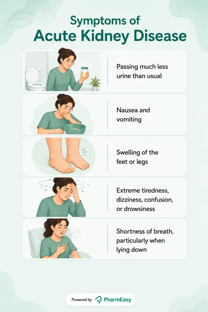
AKI develops suddenly over hours or days. A sudden drop in kidney function, suggesting AKI, would typically be associated with one or more of the following kidney disease symptoms6:
Important: AKI is a medical emergency and requires prompt evaluation and treatment. Early intervention can often restore kidney function and help avoid long-term damage4,6.
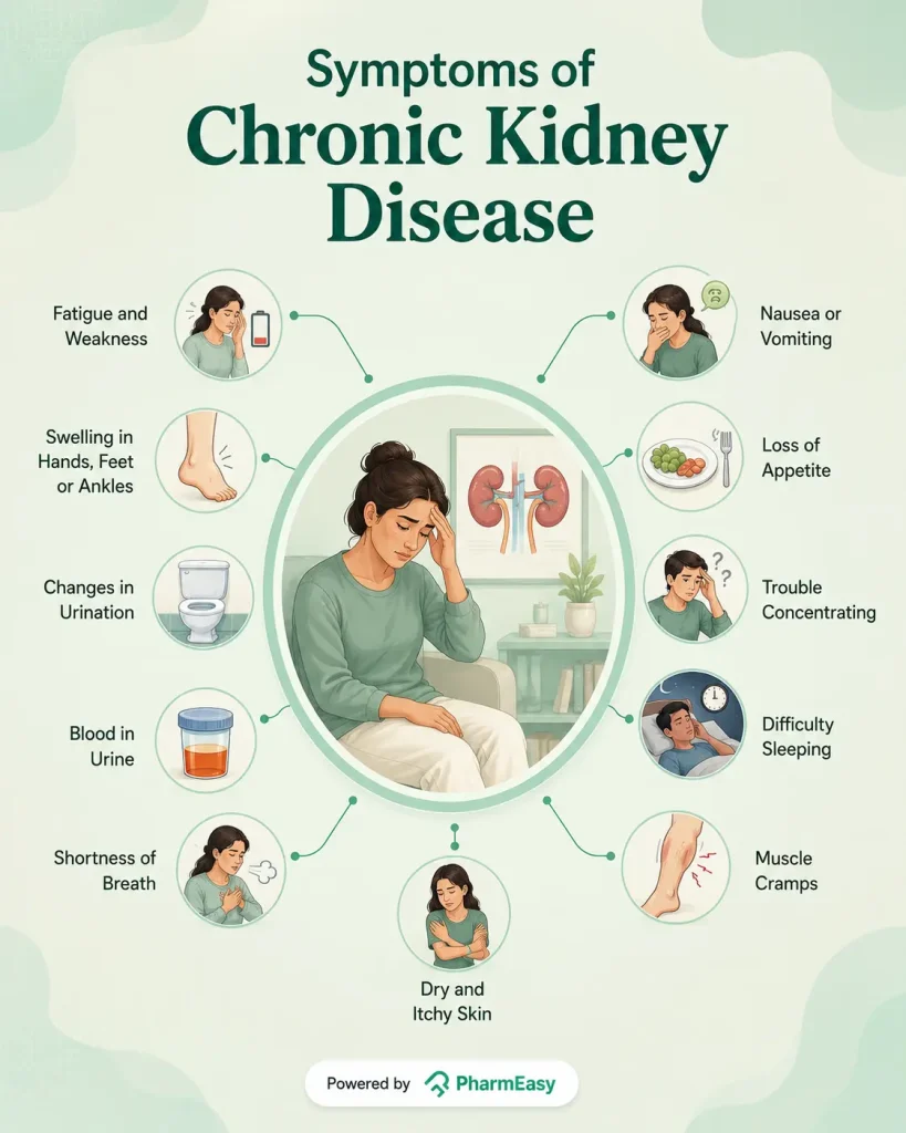
Some early signs and symptoms of CKD include:
Note: Many individuals with early CKD have no symptoms at all. Kidney disease is often detected during routine blood or urine tests performed for conditions such as diabetes or high blood pressure10.
The causes of kidney disease depend on whether it develops suddenly (AKI) or gradually (CKD).
AKI occurs when the kidneys suddenly stop working properly, often due to an illness, injury, or reduced blood flow to the kidneys. Common causes include6:
Note: The iodinated contrast dye used in some imaging tests, such as CT scans and angiography, may increase the risk of AKI in individuals with existing kidney disease or other risk factors. However, for most individuals with healthy kidneys, the risk is low6,11.
CKD develops gradually and is usually caused by long-term conditions that damage the kidneys over time. Common causes include12:
Recognising signs of kidney disease early can make a substantial difference in protecting your kidney health and overall well-being. Early detection is important for several reasons1,13:
If your doctor suspects kidney disease, they may recommend one or more of the following tests to assess how well your kidneys are working and identify the underlying cause6,14:
Note: In cases of AKI, doctors may also monitor urine output closely and perform additional tests, such as an ECG or imaging studies, depending on the suspected cause.
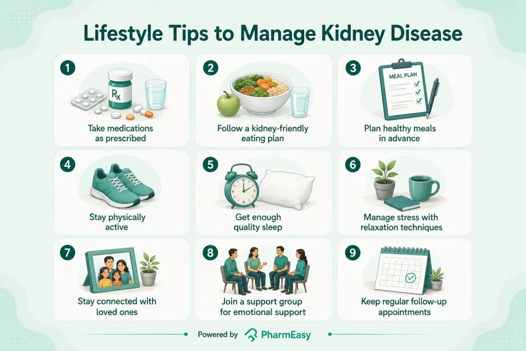
Living with kidney disease involves more than taking medications. There are several lifestyle habits that you can follow to slow disease progression, improve your quality of life, and support your overall well-being:
Also Read: Diet Chart for Kidney Patients Along with Helpful Tips
While not all kidney diseases can be prevented, healthy lifestyle habits can significantly reduce your risk. Prevention is especially important if you have diabetes, high blood pressure, heart disease, or a family history of kidney disease. Some tips for preventing kidney disease include16:
Note: Excessive water intake does not prevent CKD and can even be harmful for individuals who have been advised to restrict their fluid intake.
Also Read: High Uric Acid Level: Causes, Risks, Treatment, Prevention
Do not ignore persistent symptoms of kidney disease that could indicate a kidney problem. Early medical attention can help identify kidney disease before it becomes more serious. Consult a doctor if you6,10:
Also Read: High Creatinine: Symptoms, Causes, Diagnosis & Treatment
Kidney disease is a common but often overlooked condition that can have serious health consequences if left untreated. Therefore, knowing the early signs of kidney disease and understanding your risk can help you act before significant kidney damage occurs.
Remember, with timely medical care and healthy lifestyle choices, it is possible to protect your kidney function and maintain your overall health for years to come.
CKD is divided into five stages based on the eGFR and UACR, which measure how well the kidneys are filtering blood. Stage 1 involves mild kidney damage with normal kidney function, Stage 2 is characterised by mild to moderate loss of kidney function, Stage 3 involves moderate loss of kidney function, Stage 4 indicates severe loss of kidney function, and Stage 5 indicates kidney failure13.
Advanced kidney disease can cause unintended weight loss due to poor appetite, nausea, vomiting, and the buildup of waste products in the body. However, some individuals may initially gain weight because of fluid retention10,17.
If left untreated, kidney disease can lead to kidney failure and increase the risk of serious complications, including high blood pressure, heart disease, anaemia, and bone disorders3. Therefore, early diagnosis and management are important to help slow disease progression and reduce these risks.
1. Whaley-Connell A, Nistala R, Chaudhary K. The Importance of Early Identification of Chronic Kidney Disease. Mo Med. 2011;108(1):25-28. https://pubmed.ncbi.nlm.nih.gov/21462606/
2. Your Kidneys & How They Work – NIDDK. National Institute of Diabetes and Digestive and Kidney Diseases. Accessed July 3, 2026. https://www.niddk.nih.gov/health-information/kidney-disease/kidneys-how-they-work
3. Vaidya SR, Aeddula NR. Chronic Kidney Disease. In: StatPearls. StatPearls Publishing; 2026. Accessed July 3, 2026. http://www.ncbi.nlm.nih.gov/books/NBK535404/
4. Kidney disease. Accessed July 3, 2026. https://www.who.int/news-room/fact-sheets/detail/kidney-disease
5. Global Facts: About Kidney Disease | National Kidney Foundation. Accessed July 3, 2026. https://www.kidney.org/global-facts-about-kidney-disease
6. Acute kidney injury (AKI). nhs.uk. October 3, 2018. Accessed July 3, 2026. https://www.nhs.uk/conditions/acute-kidney-injury/
7. Khitan ZJ, Glassock RJ. Foamy Urine. Clin J Am Soc Nephrol CJASN. 2019;14(11):1664-1666. doi:10.2215/CJN.06840619 https://pubmed.ncbi.nlm.nih.gov/31575619/
8. 10 Signs You May Have Kidney Disease | National Kidney Foundation. Accessed July 3, 2026. https://www.kidney.org/news-stories/10-signs-you-may-have-kidney-disease
9. Hematuria (Blood in the Urine) – NIDDK. Accessed July 6, 2026. https://www.niddk.nih.gov/health-information/urologic-diseases/hematuria-blood-urine
10. Chronic kidney disease – Symptoms. nhs.uk. October 3, 2018. Accessed July 3, 2026. https://www.nhs.uk/conditions/kidney-disease/symptoms/
11. Wang KC, Lin LC, Pan SY, et al. Use of iodinated and gadolinium-based contrast media in patients with chronic kidney disease: Consensus statements from nephrologists, cardiologists, and radiologists at National Taiwan University Hospital. J Formos Med Assoc. 2026;125(3):261-267. doi:10.1016/j.jfma.2025.01.019 https://pubmed.ncbi.nlm.nih.gov/39870554/
12. Chronic kidney disease. nhs.uk. October 20, 2017. Accessed July 3, 2026. https://www.nhs.uk/conditions/kidney-disease/
13. KDIGO 2024 Clinical Practice Guideline for the Evaluation and Management of Chronic Kidney Disease – PubMed. Accessed July 3, 2026. https://pubmed.ncbi.nlm.nih.gov/38490803/
14. Chronic kidney disease – Diagnosis. nhs.uk. October 20, 2017. Accessed July 3, 2026. https://www.nhs.uk/conditions/kidney-disease/diagnosis/
15. 8 Self-Care Ideas for People With Chronic Kidney Disease (CKD) | National Kidney Foundation. Accessed July 3, 2026. https://www.kidney.org/news-stories/8-self-care-ideas-people-kidney-disease
16. Preventing Chronic Kidney Disease – NIDDK. National Institute of Diabetes and Digestive and Kidney Diseases. Accessed July 3, 2026. https://www.niddk.nih.gov/health-information/kidney-disease/chronic-kidney-disease-ckd/prevention
17. Singer R, Huang H. Weight change in chronic kidney disease: Association with mortality and kidney function. Obes Sci Pract. 2023;10(1):e723. doi:10.1002/osp4.723 https://pubmed.ncbi.nlm.nih.gov/38264010/
Disclaimer: The information provided here is for educational/awareness purposes only and is not intended to be a substitute for medical treatment by a healthcare professional and should not be relied upon to diagnose or treat any medical condition. The reader should consult a registered medical practitioner to determine the appropriateness of the information and before consuming any medication. PharmEasy does not provide any guarantee or warranty (express or implied) regarding the accuracy, adequacy, completeness, legality, reliability or usefulness of the information; and disclaims any liability arising thereof.
Links and product recommendations in the information provided here are advertisements of third-party products available on the website. PharmEasy does not make any representation on the accuracy or suitability of such products/services. Advertisements do not influence the editorial decisions or content. The information in this blog is subject to change without notice. The authors and administrators reserve the right to modify, add, or remove content without notification. It is your responsibility to review this disclaimer regularly for any changes.
7
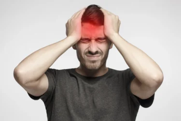
A headache is a common problem that makes it difficult to study, work, or enjoy any activities you do. Although headaches may not be severe in most cases, sometimes they may be a cause for concern. However, knowing their cause can help you get the right treatment you need1. In this blog, we’ll cover the common causes and symptoms of headaches, the available treatment options, and simple ways to help manage them.
A headache is a pain or discomfort arising from pain-sensitive structures in the head, such as the blood vessels, nerves, and the tissues covering the brain (meninges)2. This pain occurs because of changes in the way the brain, nerves, and blood vessels process pain signals. It can occur in different parts of the head, scalp, or face. It may feel dull, sharp, throbbing, or like a tight band wrapped around the head. Headaches can last from a few minutes to several hours or, in some cases, even days, depending on the underlying cause. While most headaches are not serious and improve with simple treatment, some may be a sign of an underlying health condition that requires medical attention1.
Did You Know?
Headaches are broadly classified into primary headaches and secondary headaches, depending on whether the headache itself is the main condition or is caused by another medical problem.
Headaches that occur because of changes in the pain-sensitive structures of the head or the way the brain processes pain are called primary headaches. They may not be caused by an underlying health condition. The different types of headaches (primary) include:
This is a type which indicates an underlying health condition, such as high blood pressure (BP), infection, or head injury. It can last from a few minutes to several days, weeks, or even months, depending on the underlying cause8. It happens when the condition affects the pain-sensitive tissues or nerves in the head. It is less common, but needs prompt medical evaluation and treatment for the underlying cause rather than the headache itself1. Examples include:
Other types of secondary headaches include post-traumatic headache (related to head injury or trauma), headaches related to an arterial ischemic event (caused by reduced blood flow to the brain due to a stroke), headaches related to arthritis, hypertension headache (caused by extremely high BP), and reversible cerebral vasoconstriction syndrome (caused by temporary narrowing of the blood vessels in the brain)8.
Headache symptoms can vary based on the type. While some symptoms are specific to certain types of headaches, there are a few common signs and symptoms that many people with headaches may experience. These include:
The causes of headaches vary depending on the type. Some of the most common headache causes include the following.
In addition to understanding the causes of headaches, knowing the risk factors is very important to reduce the risk or manage the condition. Factors that may increase the risk of a person having headaches include:
Note: These are common risk factors for many types of headaches. Having one or more of these risk factors does not guarantee you’ll get headaches.
The following are the approaches a doctor might take to identify the type of headache:
Headache treatment is determined by its type, severity, frequency, and underlying reason. While some headaches can be treated with simple lifestyle adjustments, others may necessitate prescription medications or therapy for the underlying medical issue. The treatment approaches that are commonly recommended include:
Depending on the type of headache, the doctor may recommend pain-relieving medicines, such as paracetamol or ibuprofen16, triptans or beta-blocker medicines (for migraine), antidepressants or barbiturates (for tension headaches; barbiturates are not routinely recommended due to a risk of medication overuse headache), verapamil (for cluster headaches)1. Although some of these medicines are available as over-the-counter medicines, taking them too often or without a doctor’s advice is not advisable12.
Headaches caused by an underlying medical condition (secondary headaches), such as sinus infections, high BP, or brain tumours, require treatment of the underlying cause. This may include antibiotics, nasal corticosteroid sprays or anti-histamines (for sinus infection)10, BP medications (for high BP), surgery (for tumours), etc.8, depending on the cause.
Exercises, stretching, and posture correction to help with tension headaches or headaches caused by muscle strain4.
Surgeries, chemotherapy, etc., in cases where the headaches do not respond to medicines, for example, a tumour that is causing a headaches5.
Note: Headache treatment differs from person to person based on the type, severity, frequency, and underlying cause. Do not self-medicate or take pain relievers on a regular basis without consulting a doctor, since this can worsen headaches.
People often wonder how to reduce headaches naturally. The best approach is to prevent headaches rather than treating them after they occur. Here are some simple ways to help reduce the risk of getting a headache:

Identifying and avoiding personal headache triggers, such as certain foods, alcohol, smoking, dehydration, bright lights, lack of sleep, improper posture, etc., can help reduce headache episodes.

Deep breathing, meditation, yoga, regular exercise and other relaxation methods can help reduce stress-related headaches1.

Undergoing regular health check-ups, especially in people with conditions such as high BP or allergies, can help identify and manage factors that may contribute to headaches.
Note: These recommendations may help reduce the frequency or severity of headaches, but they do not guarantee perfect prevention. If headaches are severe, frequent, sudden, or persistent despite these precautions, see a doctor for a correct diagnosis and treatment.
Also Read: Home Remedies For Headache By Dr. Siddharth Gupta
Consult a doctor if:
Headaches are common and, in most cases, do not indicate a major health problem. They might be caused by common conditions like stress, dehydration, lack of sleep, or illness, but they can also be symptoms of an underlying medical disease. Consult a doctor immediately for an accurate diagnosis and treatment if headaches are severe, frequent, or accompanied by strange symptoms. Understanding the various types, symptoms, and triggers might help in managing headaches more successfully.
Morning headaches can be caused by poor sleep, dehydration (mainly from alcohol consumption), stress, teeth grinding, sleep apnoea (interrupted breathing during sleep), and excessive medication use. In such cases, it is important to see a doctor to determine the underlying reason and get the right treatment17.
Headaches that worsen while bending down are commonly caused by sinus congestion or a sinus infection, as bending increases pressure in the inflamed sinuses6. However, it is important to consult a doctor for a proper diagnosis and treatment, as headaches when bending down can also be caused by other underlying conditions17.
Yes, skipped meals, especially breakfast, can result in headaches. Going too long without eating can cause blood sugar levels to drop, causing a headache, particularly in people who are prone to migraines6,17.
Yes. Dehydration may cause headaches by lowering the amount of fluid in the body,1,6 which disrupts proper brain function. Drinking plenty of water throughout the day can help avoid dehydration-related headaches. Dehydration may also occur from excessive drinking of alcohol, thereby contributing to headache17.
Yes. Lack of sleep can cause headaches by altering how your brain responds to pain. Maintaining a regular sleep schedule and getting enough rest can help lower the risk of headaches.
Headaches can happen for many reasons, including stress, lack of sleep, dehydration, skipping meals, eye strain, infections, or underlying medical conditions. Identifying the cause can help in choosing the right treatment1.
Taking a painkiller for a headache too often can actually cause more frequent headaches (medication overuse headaches). You should consult a doctor if you have recurring headaches to identify the cause rather than taking an over-the-counter medication1,12.
The best way to treat a headache depends on its cause. Resting, staying hydrated, eating a light meal if it is skipped, and taking pain-relieving medicines, if advised by a doctor or avoiding them in case of medication-overdose headaches, etc., can help relieve most common headaches1,12.
1. Headache. 2026. Available from: https://www.ninds.nih.gov/health-information/disorders/headache
2. Robertson CE, Benarroch EE. The anatomy of head pain. Handb Clin Neurol. 2023;198:41-60. doi: 10.1016/B978-0-12-823356-6.00001-9. PMID: 38043970. Available from: https://pubmed.ncbi.nlm.nih.gov/38043970/
3. Migraine and other headache disorders. 2025. Available from: https://www.who.int/news-room/fact-sheets/detail/headache-disorders
4. Nihir Shah, Asuncion RMD, Hameed S. Muscle Contraction Tension Headache. 2024. Available from: https://www.ncbi.nlm.nih.gov/books/NBK562274/
5. The Complete Headache Chart. Available from: https://headaches.org/resources/the-complete-headache-chart/
6. Headaches. 2025. Available from: https://www.healthdirect.gov.au/headaches
7. Joppeková Ľ, Pinto MJ, Da Costa MD, et al. What does a migraine aura look like?—A systematic review. J Headache Pain. 2025;26(1):149. doi:10.1186/s10194-025-02080-6 Available from: https://pubmed.ncbi.nlm.nih.gov/40597581/
8. Wijeratne T, Wijeratne C, Korajkic N, Bird S, Sales C, Riederer F. Secondary headaches – red and green flags and their significance for diagnostics. eNeurologicalSci. 2023 Jun 30;32:100473. doi: 10.1016/j.ensci.2023.100473. PMID: 37456555; PMCID: PMC10339125. Available from: https://pubmed.ncbi.nlm.nih.gov/37456555/
9. Jones NS. Sinus headaches: avoiding over- and mis-diagnosis. Expert Review of Neurotherapeutics. 2009;9(4):439-444. doi:10.1586/ern.09.8 Available from: https://pubmed.ncbi.nlm.nih.gov/19344297/
10. Sinusitis. 2024. Available from: https://medlineplus.gov/ency/article/000647.htm
11. How to Know if You Have Migraine or Sinus Headache. 2023. Available from: https://americanmigrainefoundation.org/resource-library/sinus-headache/
12. Medication Overuse Headache. 2022. Available from: https://headaches.org/resources/medication-overuse-headache/
13. Aggarwal M, Puri V, Puri S. Serotonin and CGRP in migraine. Ann Neurosci. 2012 Apr;19(2):88-94. doi: 10.5214/ans.0972.7531.12190210. PMID: 25205974; PMCID: PMC4117050. Available from: https://pubmed.ncbi.nlm.nih.gov/25205974/
14. Sinus Infection Basics. 2024. Available from: https://www.cdc.gov/sinus-infection/about/index.html
15. Kim KT. Lumbar puncture: considerations, procedure, and complications. encephalitis. 2022;2(4):93-97. doi:10.47936/encephalitis.2022.00045 Available from: https://pubmed.ncbi.nlm.nih.gov/37469996/
16. Medicines for headaches. 2025. Available from: https://www.healthdirect.gov.au/medicines-for-headaches
17. Hong Y, Kang MK, Kim MS, Mo H, Cox RC, Im HJ. Morning Headaches: An In-depth Review of Causes, Associated Disorders, and Management Strategies. Headache and Pain Research. 2025;26(1):66-79. doi:10.62087/hpr.2024.0023 Available from: https://www.researchgate.net/publication/405443332_Comments_on_Morning_Headaches_An_In-Depth_Review_of_Causes_Associated_Disorders_and_Management_Strategies
Disclaimer: The information provided here is for educational/awareness purposes only and is not intended to be a substitute for medical treatment by a healthcare professional and should not be relied upon to diagnose or treat any medical condition. The reader should consult a registered medical practitioner to determine the appropriateness of the information and before consuming any medication. PharmEasy does not provide any guarantee or warranty (express or implied) regarding the accuracy, adequacy, completeness, legality, reliability or usefulness of the information; and disclaims any liability arising thereof.
Links and product recommendations in the information provided here are advertisements of third-party products available on the website. PharmEasy does not make any representation on the accuracy or suitability of such products/services. Advertisements do not influence the editorial decisions or content. The information in this blog is subject to change without notice. The authors and administrators reserve the right to modify, add, or remove content without notification. It is your responsibility to review this disclaimer regularly for any changes.
You got your blood test back, and creatinine is flagged. You are not sure what it means or whether to worry. Most people have never heard of creatinine until a report puts it in front of them. The good news is that a high reading does not always mean something serious. But it does deserve attention. In this blog, we will understand high creatinine meaning, causes, possible high creatinine symptoms, and treatment options to manage the effects of high creatinine.
Creatinine is a waste product, usually produced during the breakdown of muscles. Healthy kidneys filter creatinine out of the blood through the urine. Traces of some creatinine can be found in everyone; however, it becomes concerning when creatinine levels are high1.
High creatinine levels may indicate impaired kidney function, while low levels are usually associated with low muscle mass, poor nutrition, or certain medical conditions and rarely indicate kidney disease2.
Creatinine levels are assessed through a blood test and expressed in milligrams per decilitre (mg/dL). The normal range can vary depending on factors such as age, sex, and muscle mass3. Generally, the following values are considered normal for creatinine:
Serum creatinine levels may show minor variations depending on factors such as the time of the day, time in the menstrual cycle, and dietary intake. Very high or persistently high creatinine levels may be associated with abnormal kidney function4. Creatinine levels above 1.1 mg/dL in females and above 1.3 mg/dL in males are considered high. Let’s understand the causes of high creatinine levels.
There are several causes of high creatinine levels, ranging from eating certain foods to conditions like underlying kidney disorders. Reasons for high creatinine levels include:
High creatinine levels may not cause any symptoms in some individuals and may only be detected through blood tests. When symptoms do occur, they are usually due to the underlying condition affecting the kidneys and not the direct effects of high creatinine or signs of high creatinine itself. These include:2
These symptoms can vary depending on the underlying cause and the severity of kidney impairment.
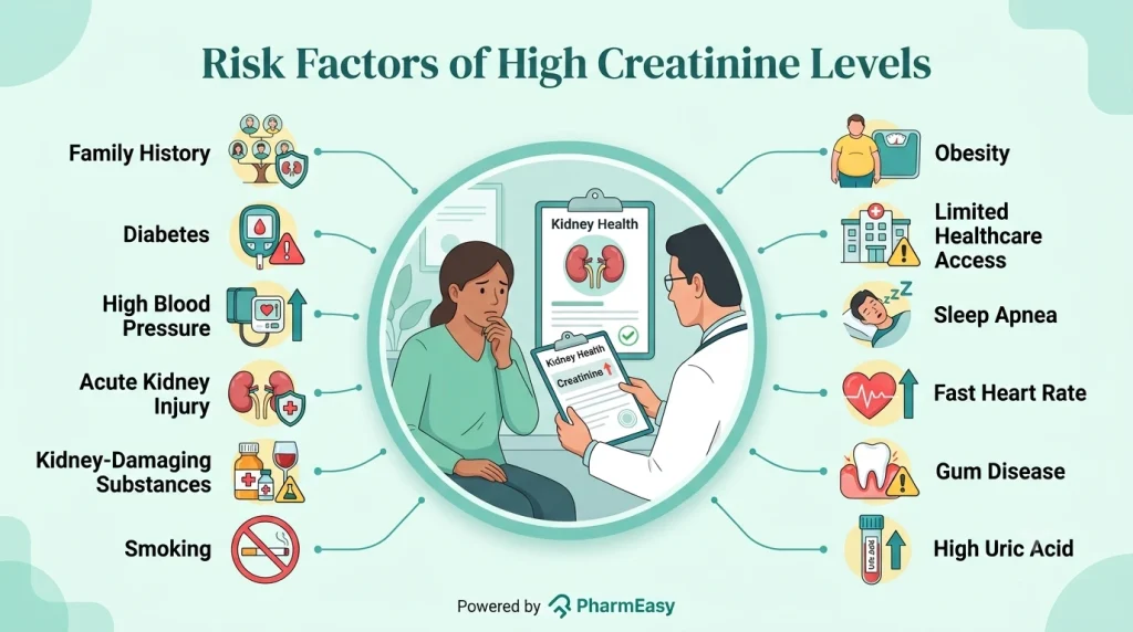
High creatinine levels are commonly associated with kidney damage or reduced kidney function. The following health conditions and lifestyle factors may increase the risk of developing elevated creatinine levels:
High creatinine levels can result from a variety of conditions. Diagnosing the underlying condition may need a combination of blood tests, urine tests, and, in some cases, imaging studies to get a clear picture of any health issues. Common diagnostic tests for conditions associated with high creatinine levels may include:
Creatinine is usually a marker and not a disease. Therefore, the treatment is directed towards the underlying cause that may have led to the high creatinine levels. Depending on the lab reports, treatment may involve:
One may be able to avoid high creatinine levels by effectively managing conditions that may damage the kidneys, such as diabetes and high blood pressure. Some simple steps could be:

This is one of the most effective ways of protecting kidney function and delaying the onset of kidney disease. The blood pressure goal should be less than 130/80 mmHg or as discussed with your doctor. Eating healthy, quitting smoking, having an active lifestyle, and getting adequate sleep are some ways that can help to maintain the blood pressure10.

Regular monitoring of blood glucose levels can help guide decisions about diet, medications, and physical activity. This allows better day-to-day diabetes management. Keeping blood glucose and HbA1c levels within the target range can help reduce the risk of diabetic kidney disease10.

Regular kidney function tests are important not only for diagnosing kidney disease in individuals at risk of kidney disease but also for monitoring its progression over time. Since kidney disease often progresses gradually, eGFR and urine albumin levels help to track changes in the kidneys10.

Some over-the-counter (OTC) and prescription medications, particularly non-steroidal anti-inflammatory drugs (NSAIDs), can affect kidney function if used for prolonged periods. These medicines are commonly found in products used to relieve pain, fever, headaches, and cold symptoms. Before using pain relievers regularly, it is advisable to consult a doctor10.
Diet plays an important role in maintaining kidney health. A balanced, kidney-friendly eating plan can help support kidney function, manage blood pressure and blood sugar levels, and reduce the risk of complications associated with kidney disease10.

About 30 minutes of moderate-intensity physical activity a day can help regulate blood sugar levels, support cardiovascular health, and aid in weight management10.
Cigarette smoking may be associated with impaired kidney function. Quitting smoking may help to maintain blood pressure, which in turn is good for the kidneys and overall health10.

This is important for overall health. Adequate sleep can help regulate blood pressure, improve blood sugar levels, and contribute to better physical and mental well-being10.

Chronic stress can negatively affect overall health by contributing to high blood pressure, poor blood sugar control, and other factors that may impact kidney function. Finding healthy ways to manage stress can support both physical and emotional well-being10.
Also Read: Kidney Failure: Symptoms, Causes, Treatment & Prevention
While mildly elevated creatinine may resolve with hydration or dietary adjustments, certain symptoms require prompt medical attention. You should visit a doctor if you have:
Creatinine levels are an important indicator of kidney health and can provide valuable insights into kidney function. While high creatinine levels may occur due to factors such as dehydration or taking certain medicines, they may also indicate kidney conditions. Early detection and management of these issues can help the management of these conditions and reduce the risk of complications. Regular health checkups, healthy lifestyle habits, and timely treatment of conditions such as diabetes and high blood pressure can go a long way in protecting kidney function.
High creatinine levels may indicate that the kidneys are not functioning effectively. While mildly elevated creatinine levels may be reversible, high levels may indicate conditions such as CKD and AKI1.
A creatinine level above 1.3 mg/dL in men or 1.1 mg/dL in women may be higher than the normal range and could indicate reduced kidney function. This normal range may vary between laboratories. As creatinine levels vary based on factors such as age, muscle mass, medical history, and changes in creatinine levels over time, they should be interpreted with the help of a doctor3.
If dehydration is the cause of elevated creatinine, adequate hydration may help normalise creatinine levels1.
In many cases, yes. If the cause of high creatinine levels is dehydration, medication use, or a temporary condition such as AKI, creatinine levels may return to normal with appropriate management. If elevated creatinine levels are caused by CKD, treatment usually focuses more on slowing the progression of CKD rather than reversing high creatinine levels1,2.
Yes, protein shakes may temporarily increase creatinine levels in some individuals, however, it may not indicate kidney disease in healthy individuals. However, in individuals with kidney disease, high protein intake can accelerate kidney damage1.
Yes, kidney stones may cause high creatinine levels. Treating kidney stones may help improve creatinine levels11.
Disclaimer: The information provided here is for educational/awareness purposes only and is not intended to be a substitute for medical treatment by a healthcare professional and should not be relied upon to diagnose or treat any medical condition. The reader should consult a registered medical practitioner to determine the appropriateness of the information and before consuming any medication. PharmEasy does not provide any guarantee or warranty (express or implied) regarding the accuracy, adequacy, completeness, legality, reliability or usefulness of the information; and disclaims any liability arising thereof.
Links and product recommendations in the information provided here are advertisements of third-party products available on the website. PharmEasy does not make any representation on the accuracy or suitability of such products/services. Advertisements do not influence the editorial decisions or content. The information in this blog is subject to change without notice. The authors and administrators reserve the right to modify, add, or remove content without notification. It is your responsibility to review this disclaimer regularly for any changes.
Tuberculosis (TB) is a contagious infection caused by the bacteria Mycobacterium tuberculosis. It primarily affects the lungs (pulmonary TB) but can spread to other parts of the body, such as the bones, spine, or brain1. This article will explain what bone tuberculosis is, its forms, common symptoms, and how to manage it efficiently. Early detection of this disease is critical since timely diagnosis and treatment can reduce long-term problems and enhance the chances of recovery.
Bone tuberculosis is a form of TB that occurs when the infection occurs in the bones or joints. It is also known as skeletal tuberculosis or osteoarticular tuberculosis. TB of the spine is the most common type of bone TB, also known as Pott’s disease. Other commonly affected regions include the hip, knee, ankle, and long bones. It develops slowly, may go unnoticed in the early stages, and can cause significant bone destruction over time if not diagnosed and treated early2.
TB can affect different parts of the skeletal system. The different types of bone TB include:

This is the most common type of bone tuberculosis, which affects the spinal vertebrae. It can cause the spinal bones to collapse, resulting in severe back pain and deformities such as kyphosis (hunchback). In severe disease, it can compress the spinal cord, resulting in neurological symptoms such as numbness of the extremities and buttock area and weakness3.

This type affects the long weight-bearing bone joints, such as the knees, hips, foot, and ankle. It develops slowly and leads to chronic pain, swelling, and stiffness in the affected joint. Over time, it can destroy joint cartilage and reduce mobility significantly. If untreated, it may result in permanent joint deformity4.

This one can affect almost any bone in the body, including the ribs, skull, pelvis, and long bones. It often develops through more than one route of infection5. In children, especially aged under 6, it can affect even fingers and toes (known as spina ventosa)6.

This condition affects more than one bone or joint at the same time or one after another. Commonly affected areas include the spine, pelvis, hip (femoral head), shoulders, and knees. It can cause symptoms like joint pain, stiffness, swelling, redness, warmth over the area, and difficulty in moving the joint. In severe cases, changes in bone shape or deformity may occur7.
Also Read: Spinal Tuberculosis (Pott’s Disease): Symptoms, Treatment, Diagnosis & More
Bone TB may be caused by:
Bone tuberculosis symptoms may develop gradually and can vary depending on the affected bone or joint. They can be divided into two:
Bone TB rarely causes general symptoms affecting the whole body. However, some people may experience the following:
Bone-related symptoms may occur due to damage and inflammation in the affected bones or joints. These include:
Bone TB is not usually communicable. Unlike pulmonary TB, which affects the lungs and can spread through the air when an infected person coughs, sneezes, or speaks1, bone TB develops when the TB bacteria travel from another region of the body to the bones or joints2,5.
A person with bone TB cannot spread the infection to others by physical contact, touching, sharing food, or being near someone. As a result, patients with bone TB are typically not considered contagious. However, if a person has both bone TB and active pulmonary TB, they may spread the TB bacteria through respiratory droplets from their lungs. In such cases, the lung infection is responsible for the transmission of TB, not the bone infection.
Bone tuberculosis diagnosis can be difficult since its symptoms usually appear gradually and resemble those of other bone and joint problems. Doctors usually confirm the diagnosis with a medical history, physical examination, imaging studies, and laboratory investigations.
The doctor will ask about symptoms such as prolonged bone or joint pain, swelling, stiffness, fever, weight loss, and any previous TB exposure. They might also look for neurological symptoms (weakness, numbness, etc.), restricted joint movement, discomfort, and other signs and symptoms2.
The doctor might recommend blood tests such as:
The doctor may recommend this test to check for possible TB infection or exposure. The test checks for a skin reaction after injecting a small amount of tuberculin solution, usually on the forearm1,4.
The doctor might recommend some imaging tests to get a detailed view of the bones and joints:
To confirm the diagnosis of bone TB, the doctor may do a biopsy, which involves taking a small sample of tissue from the diseased bone and examining it. The sample is then sent to the laboratory to confirm the presence of the bacteria using tests2 such as AFB culture, which can help detect active TB infection and enable correct treatment planning. Please visit the site below to know more about the AFB (MGIT) test
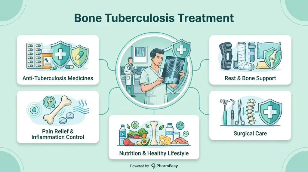
Bone tuberculosis treatment primarily focuses on eliminating the TB bacteria, reducing pain and inflammation, and avoiding long-term damage to bones and joints. The doctor might recommend the following as treatment approaches:
If bone tuberculosis is not treated promptly, it can slowly progress and cause a variety of complications, such as:
Recovery from bone or spinal TB is usually slow and depends on how early the disease is detected and how well treatment is taken6. Most patients require several months of consistent medication, and improvement occurs gradually over time3,8 Pain and other symptoms usually begin to subside within a few weeks to months, although complete healing of bones and joints may take longer.
Along with completing the full course of TB treatment, following the lifestyle changes listed below may help improve the effectiveness of treatment:
Consult a doctor immediately for the following:
Also Read: Tuberculosis: Types, Causes, Treatment and Prevention
Bone tuberculosis is a serious but treatable form of TB that affects the bones and joints, most commonly the spine. Because its symptoms often appear gradually, early detection is critical for avoiding problems like bone degeneration, joint deformity, and nerve damage. Most patients can recover successfully if they receive immediate treatment, which includes antitubercular drugs and appropriate supportive care. Following the treatment as prescribed, eating a healthy diet, and attending regular doctor visits can help support recovery and lower the risk of complications.
People with persistent bone, joint, or back pain, swelling, stiffness, trouble moving the affected area, and symptoms such as fever, weight loss, or night sweats should be checked for bone TB. It may be diagnosed with the help of imaging tests (X-ray, CT scan, or MRI) and confirmed by tests, most commonly a bone or tissue sample4.
Yes, bone TB can usually be cured with early detection and a complete course of anti-tubercular drugs. Early treatment reduces problems and increases the likelihood of a full recovery3.
Bone TB can be avoided by lowering the risk of TB infection through early detection and treatment of active TB, avoiding close contact with untreated TB patients, and keeping a strong immune system. Good nutrition, good hygiene, and well-ventilated living areas may help reduce the risk of TB1,2.
Processed and junk foods, extra sugar, and alcohol11 should be avoided during bone TB treatment since they may decrease immunity and slow healing. Limit the intake of particularly fatty or deep-fried foods, as these may interfere with general healing and nourishment.
1. Tobin EH, Tristram D. Tuberculosis Overview. 2024. https://www.ncbi.nlm.nih.gov/books/NBK441916/
2. Pigrau-Serrallach C, Rodríguez-Pardo D. Bone and joint tuberculosis. Eur Spine J. 2013;22(S4):556-566. doi:10.1007/s00586-012-2331-y https://pubmed.ncbi.nlm.nih.gov/22711012/
3. Tobin EH, Rausch-Phung EA. Tuberculous Spondylitis (Pott Disease). 2026. https://www.ncbi.nlm.nih.gov/books/NBK538331/
4. Marais LC, Nieuwoudt L, Nansook A, Menon A, Benito N. Tuberculous arthritis of native joints – a systematic review and European Bone and Joint Infection Society workgroup report. J Bone Joint Infect. 2023;8(4):189-207. doi:10.5194/jbji-8-189-2023 https://pubmed.ncbi.nlm.nih.gov/37780528/
5. Emerson P, Philip A, Varghese GM, Thomas R. Tuberculous Osteomyelitis of the Hyoid Bone: A Case Report. Case Reports in Otolaryngology. 2013;2013:1-3. doi:10.1155/2013/549564 https://pmc.ncbi.nlm.nih.gov/articles/PMC3610351/
6. N. AlGhazi A, H. AlZahrani M, AlMutiri WA, AlZoum NM. Disseminated tuberculosis presenting as finger swelling in a 2-year-old: a case report of TB osteomyelitis. Case Reports in Plastic Surgery and Hand Surgery. 2025;12(1):2473383. doi:10.1080/23320885.2025.2473383 https://pmc.ncbi.nlm.nih.gov/articles/PMC11899214/
7. Zhou J, Yang X, Hu Y, Li S. Epidemiological and osteoarticular involvement sites’ characteristics of multiple osteoarticular tuberculosis: a scoping review. Epidemiol Infect. 2025;153:e26. doi:10.1017/S095026882400150X https://pubmed.ncbi.nlm.nih.gov/39834064/
8. Herdea A, Marie H, Negrila IA, Abdel Hamid Ahmed AD, Ulici A. Reevaluating Pediatric Osteomyelitis with Osteoarticular Tuberculosis: Addressing Diagnostic Delays and Improving Treatment Outcomes. Children. 2024;11(11):1279. doi:10.3390/children11111279 https://pubmed.ncbi.nlm.nih.gov/39594854/
9. Gupta K, Gupta R, Atreja A, Verma M, Vishvkarma S. Tuberculosis and nutrition. Lung India. 2009;26(1):9. doi:10.4103/0970-2113.45198 https://pubmed.ncbi.nlm.nih.gov/20165588/
10. Spinal Tuberculosis: Rural India’s Hidden Epidemic. https://thespinefoundation.org/spinal-tuberculosis-hidden-epidemic/
11. Heshmati B, Omidi S, Mohammadi Y. Impact of alcohol consumption, substance use, and smoking on treatment outcomes in tuberculosis: a systematic review and meta-analysis. Syst Rev. 2025;14(1):139. doi:10.1186/s13643-025-02888-y https://pubmed.ncbi.nlm.nih.gov/40618124/
Disclaimer: The information provided here is for educational/awareness purposes only and is not intended to be a substitute for medical treatment by a healthcare professional and should not be relied upon to diagnose or treat any medical condition. The reader should consult a registered medical practitioner to determine the appropriateness of the information and before consuming any medication. PharmEasy does not provide any guarantee or warranty (express or implied) regarding the accuracy, adequacy, completeness, legality, reliability or usefulness of the information; and disclaims any liability arising thereof.
Links and product recommendations in the information provided here are advertisements of third-party products available on the website. PharmEasy does not make any representation on the accuracy or suitability of such products/services. Advertisements do not influence the editorial decisions or content. The information in this blog is subject to change without notice. The authors and administrators reserve the right to modify, add, or remove content without notification. It is your responsibility to review this disclaimer regularly for any changes.
Many people who use insulin every day follow their routine with confidence, yet small, unnoticed mistakes can sometimes make it harder to keep blood sugar levels under control. It could be something as simple as incorrect storage, using the wrong injection technique, or missing the right dose timing that can affect how well insulin works1,2.
The good news is that these mistakes are common and often easy to correct. In this article, we’ll explore the most common insulin injection mistakes, why they matter, and practical tips to help you use insulin safely and effectively.
Insulin is a life-saving medicine for people with type 1 diabetes and for many people with type 2 diabetes1. It helps keep blood sugar levels within the target range, thereby reducing both high and low blood sugar cases.
However, insulin can only work effectively when it is used correctly. Below are the reasons why proper usage matters.
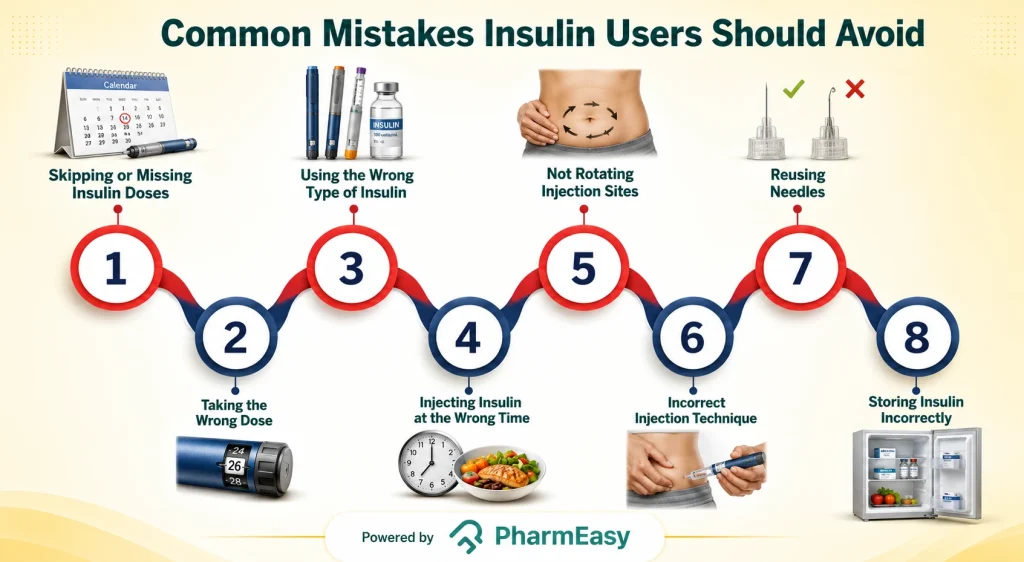
Even experienced insulin injection users can make small mistakes that could affect blood sugar control. Therefore, being aware of these common errors can help you get the most benefit from your insulin and reduce the risk of complications. Some common mistakes include:
Proper storage helps insulin remain effective and safe to use. Follow these practical tips to avoid common storage mistakes1,6:
Using an insulin injection can sometimes be challenging, but a few practical steps can help you manage common issues and improve blood sugar control. Here’s what you can do:

Along with taking insulin correctly, healthy daily habits can also help improve blood sugar control and overall diabetes management. Here are some to follow:
Missing an insulin dose may happen accidentally. What you should do next depends on the type of insulin you use, when the missed dose is noticed, and your current blood sugar level.
If you miss or forget an insulin dose11,13:
Tip: Set reminders using alarms, smartphone apps, or pill and medication reminder tools to help avoid missed doses in the future.
If your insulin is not working as expected, your blood sugar levels may become difficult to control. Some signs may include:
Like all medicines, insulin may cause side effects, although not everyone experiences them. Common insulin side effects include14:
Important: Severe allergic reactions, including rash, swelling, or difficulty breathing, may rarely occur. These require immediate medical attention.
Contact your doctor if you experience any of the following:
Note: Whenever you have concerns about your insulin therapy or blood sugar control, never stop or adjust your insulin without consulting your doctor.
Also Read: Insulin Resistance: What You Need To Know
Insulin works best when it is used correctly and consistently. Try to follow simple steps, such as storing insulin properly, using the correct injection technique, rotating injection sites, monitoring your blood sugar, and following your prescribed dose, which can help insulin work effectively and improve blood sugar control.
Also, you should learn to recognise the signs of low and high blood sugar and never hesitate to contact your doctor if you have concerns or notice persistent changes in your blood sugar levels.
No, it is recommended to use a new needle for every injection. Reusing needles can make injections more painful, increase the risk of infection, damage the needle tip, and contribute to thickened skin, which could affect insulin absorption16.
Unopened insulin should be stored according to the manufacturer’s instructions, usually in a refrigerator (2°C to 8°C) until its expiry date. Once in use, most insulin products can be kept at room temperature (generally below 30°C) for a limited period, which varies by product1,6. Always follow the storage instructions provided with your specific insulin.
Insulin may not work properly if it is expired, stored incorrectly, frozen, exposed to excessive heat, injected into thickened or damaged skin, administered using poor injection technique, or if the wrong dose or type of insulin is used1,2.
Rotating injection sites helps prevent lumps or thickened skin, promotes more consistent insulin absorption, and improves blood sugar control. The recommended injection sites include the abdomen, thighs, upper arms, and buttocks. You should also avoid injecting into the same spot repeatedly6.
1. Tulsan SK, Laila R, Patel H, et al. Errors in diabetic insulin therapy and the vitality of proper precautions in Bangladesh: Real-life insights from the developing world. J Fam Med Prim Care. 2024;13(1):292-297. doi:10.4103/jfmpc.jfmpc_484_23. https://pubmed.ncbi.nlm.nih.gov/38482322/
2. Trief PM, Cibula D, Rodriguez E, Akel B, Weinstock RS. Incorrect Insulin Administration: A Problem That Warrants Attention. Clin Diabetes Publ Am Diabetes Assoc. 2016;34(1):25-33. doi:10.2337/diaclin.34.1.25. https://pubmed.ncbi.nlm.nih.gov/26807006/
3. Services D of H& H. Diabetes and insulin. Accessed June 25, 2026. http://www.betterhealth.vic.gov.au/health/conditionsandtreatments/diabetes-and-insulin
4. Home P, Riddle M, Cefalu WT, et al. Insulin Therapy in People With Type 2 Diabetes: Opportunities and Challenges? Diabetes Care. 2014;37(6):1499-1508. doi:10.2337/dc13-2743. https://pubmed.ncbi.nlm.nih.gov/24855154/
5. Rahman MS, Hossain KS, Das S, et al. Role of Insulin in Health and Disease: An Update. Int J Mol Sci. 2021;22(12):6403. doi:10.3390/ijms22126403. https://pubmed.ncbi.nlm.nih.gov/34203830/
6. Tandon N, Kalra S, Balhara YPS, et al. Forum for Injection Technique (FIT), India: The Indian recommendations 2.0, for best practice in Insulin Injection Technique, 2015. Indian J Endocrinol Metab. 2015;19(3):317-331. doi:10.4103/2230-8210.152762. https://pmc.ncbi.nlm.nih.gov/articles/PMC4366768/
7. Signs, Symptoms, and Treatment for Hypoglycemia (Low Blood Glucose) | American Diabetes Association. Accessed June 25, 2026. https://diabetes.org/living-with-diabetes/hypoglycemia-low-blood-glucose/symptoms-treatment
8. Hyperglycemia (High Blood Glucose) | American Diabetes Association. Accessed June 25, 2026. https://diabetes.org/living-with-diabetes/treatment-care/hyperglycemia
9. Apr 4 LR, 2024. Living Healthy with Diabetes. www.heart.org. Accessed June 25, 2026. https://www.heart.org/en/health-topics/diabetes/prevention–treatment-of-diabetes/living-healthy-with-diabetes
10. Chacko E, Signore C. Five Evidence-Based Lifestyle Habits People With Diabetes Can Use. Clin Diabetes Publ Am Diabetes Assoc. 2020;38(3):273-284. doi:10.2337/cd19-0078. https://pmc.ncbi.nlm.nih.gov/articles/PMC7364446/
11. How and when to take long-acting insulin. nhs.uk. July 11, 2023. Accessed June 25, 2026. https://www.nhs.uk/medicines/insulin/long-acting-insulin/how-and-when-to-take-long-acting-insulin/
12. How and when to take short-acting insulin. nhs.uk. July 11, 2023. Accessed June 25, 2026. https://www.nhs.uk/medicines/insulin/short-acting-insulin/how-and-when-to-take-short-acting-insulin/
13. How and when to take intermediate-acting insulin. nhs.uk. July 11, 2023. Accessed June 25, 2026. https://www.nhs.uk/medicines/insulin/intermediate-acting-insulin/how-and-when-to-take-intermediate-acting-insulin/
14. Side effects of long-acting insulin. nhs.uk. July 11, 2023. Accessed June 25, 2026. https://www.nhs.uk/medicines/insulin/long-acting-insulin/side-effects-of-long-acting-insulin/
15. Diabetes and being ill | Managing when you’re sick | Diabetes UK. Accessed June 25, 2026. https://www.diabetes.org.uk/living-with-diabetes/life-with-diabetes/illness
16. Impact of Insulin Injection and Infusion Routines on Lipohypertrophy and Glycemic Control in Children and Adults with Diabetes – PMC. Accessed June 25, 2026. https://pmc.ncbi.nlm.nih.gov/articles/PMC6349294/
Disclaimer: The information provided here is for educational/awareness purposes only and is not intended to be a substitute for medical treatment by a healthcare professional and should not be relied upon to diagnose or treat any medical condition. The reader should consult a registered medical practitioner to determine the appropriateness of the information and before consuming any medication. PharmEasy does not provide any guarantee or warranty (express or implied) regarding the accuracy, adequacy, completeness, legality, reliability or usefulness of the information; and disclaims any liability arising thereof.
Links and product recommendations in the information provided here are advertisements of third-party products available on the website. PharmEasy does not make any representation on the accuracy or suitability of such products/services. Advertisements do not influence the editorial decisions or content. The information in this blog is subject to change without notice. The authors and administrators reserve the right to modify, add, or remove content without notification. It is your responsibility to review this disclaimer regularly for any changes.
Most of us have heard people talk about the dangers of high cholesterol. But have you heard about problems caused by low cholesterol? Yes, that can happen too, especially with low high-density lipoprotein (HDL), often called the good cholesterol, and yes, it deserves attention!
Cholesterol is a fatty substance that is essential for many important functions in the body. It helps maintain the structure and flexibility of cell membranes. It also plays a key role in producing vitamin D, hormones such as cortisol and testosterone, and bile acids that aid digestion and the absorption of vitamins A, D, E, and K. Since cholesterol does not dissolve easily in blood, it is transported through the bloodstream by lipoproteins (biochemical molecules made of protein and fat) such as HDL, low-density lipoprotein (LDL), and very-low-density lipoprotein (VLDL), which together contribute to total cholesterol levels. Abnormal levels of any of these lipoproteins can lead to health problems, particularly those related to heart health1.
In this blog, we will explore what HDL cholesterol low means, its causes, symptoms, risks, and effective ways to increase it naturally and maintain better heart health.
As discussed, cholesterol travels through the bloodstream with the help of lipoproteins. Among these, LDL and HDL are the two main types.
The following table provides the average values for optimal HDL cholesterol levels4.
| Group | Healthy HDL Level | Low HDL Level |
| Men (Age 20 or Older) | Greater than or equal to 60 mg/dL | Less than 40 mg/dL |
| Women (Age 20 or Older) | Greater than or equal to 60 mg/dL | Less than 50 mg/dL |
Note: Results may vary from person to person and should always be interpreted by a doctor.
Low HDL cholesterol means the body is becoming less effective at clearing excess cholesterol from the bloodstream. This can increase the risk of plaque buildup in the arteries, coronary artery disease, heart attack, and stroke5.
Important: Low HDL is considered an independent risk factor for cardiovascular disease5.
HDL cholesterol low usually does not cause noticeable symptoms in most people and is often detected during a routine blood test. However, certain underlying conditions associated with low HDL may show physical signs that can indicate an increased cardiovascular risk.
Some low HDL cholesterol symptoms include6:
The answer to why HDL cholesterol is low can lie in several medical, genetic, and medication-related factors. Certain low HDL cholesterol causes include6:
Some inherited disorders affect cholesterol metabolism and lead to very low HDL levels. These include6:
Note: Certain medications may also contribute to low HDL cholesterol levels, including beta blockers and anabolic steroids.
Many lifestyle factors can affect HDL cholesterol levels in the body. Common risk factors of low HDL cholesterol include6:
Low HDL cholesterol is usually diagnosed through a fasting lipid profile blood test, which measures6:
In some cases, additional tests such as nuclear magnetic resonance (NMR) may be used to measure lipoprotein particle numbers, although these tests are not routinely used.
Doctors may also recommend other investigations to identify underlying causes associated with low HDL cholesterol. These may include6:
Note: Although these signs may occur in some cases, HDL cholesterol low itself is usually ‘silent.’ Regular cholesterol screening is therefore important for early detection and management.
There is currently no specific treatment recommended solely to increase HDL cholesterol levels. Instead, management mainly focuses on reducing overall cardiovascular risk and improving heart health through lifestyle changes.
However, some cholesterol-lowering medications, such as statins, may slightly increase HDL levels while effectively lowering LDL cholesterol. Another medication called niacin might significantly raise HDL cholesterol levels; however, studies have shown limited additional cardiovascular benefit when added to standard statin therapy6.
Important: Do not self-medicate or start any cholesterol-lowering medicines or supplements without consulting a doctor. Proper medical evaluation and guidance are important to determine the underlying cause of low HDL cholesterol and the most appropriate treatment approach.
Making healthy lifestyle changes can help improve HDL cholesterol levels and support overall heart health. Some effective ways to increase HDL cholesterol include2:
Choose healthy unsaturated fats over saturated and trans fats. We will discuss more dietary options in the next section.
Losing excess weight (especially around the waist) can help raise HDL cholesterol levels and reduce cardiovascular risk.
Regular physical activity, especially aerobic exercises such as walking, jogging, cycling, or swimming, can help increase HDL cholesterol while lowering LDL (bad) cholesterol. Aim for at least 30 minutes of exercise on most days of the week.
Smoking lowers HDL cholesterol levels and damages blood vessels. Quitting smoking can improve HDL levels and overall heart health.
Reducing sugary foods, sweetened beverages, and refined carbohydrates may help improve HDL cholesterol levels.
Excessive alcohol consumption can contribute to weight gain and negatively affect cholesterol levels. So, alcohol should only be consumed in moderation, if at all.
Including heart-healthy foods in your daily diet may help improve HDL cholesterol levels and support overall cardiovascular health. Some beneficial food options include:
At the same time, it is important to limit fried foods, processed snacks, sugary foods, trans fats, and excessive saturated fats, as these may negatively affect cholesterol levels and heart health2.
Low HDL cholesterol can increase the risk of several health complications, particularly those related to heart and blood vessel health. Possible complications include6:
Regular health checkups and lipid profile testing can help detect low HDL cholesterol early and reduce the risk of future heart-related complications. You should consult a doctor if you:
Also Read: High LDL (Bad) Cholesterol: Causes, Symptoms & How to Reduce It
HDL, or good cholesterol, plays an important role in protecting heart health by helping remove excess cholesterol from the bloodstream. Low HDL cholesterol levels may increase the risk of heart disease, stroke, and other metabolic complications, especially when combined with unhealthy lifestyle habits or underlying medical conditions.
Although low HDL often does not cause noticeable symptoms, regular cholesterol screening can help with early detection and management. Further, adopting healthy lifestyle habits such as eating a balanced diet, exercising regularly, maintaining a healthy weight, and avoiding smoking can help improve HDL levels and support overall cardiovascular health.
If you have persistently low HDL cholesterol or other risk factors for heart disease, consult a doctor for proper evaluation, diagnosis, and personalised treatment guidance.
HDL cholesterol usually improves with diet. This may support better HDL levels over time2.
Some drinks, such as green tea, have shown benefits in helping reduce LDL (bad) cholesterol levels, although their effect on raising HDL cholesterol is not very significant10. Thus, focusing on an overall healthy lifestyle, balanced diet, regular exercise, and weight management is more important for improving HDL cholesterol levels and supporting heart health
Yes. Low HDL cholesterol can increase the risk of plaque buildup in the arteries, heart disease, stroke, and other cardiovascular complications, especially when combined with high LDL cholesterol or other risk factors6.
Improving HDL cholesterol levels depends on lifestyle changes, diet, exercise habits, weight management, and overall health. A doctor can help guide this process based on your individual medical history, risk factors, and test results.
In general, higher HDL (‘good’) cholesterol levels are considered better for heart health because HDL helps remove excess cholesterol from the bloodstream. Low HDL levels may increase the risk of heart disease and stroke3,6. However, cholesterol levels should always be interpreted in the context of your overall health, medical history, and other lipid levels. You should always consult a doctor for proper evaluation and personalised guidance.
Fruits rich in fibre and antioxidants may help support healthy HDL levels. These include apple, grapes, mango, pineapple, watermelon, orange, kiwi, pomegranate, avocado, and papaya, as part of a balanced, heart-healthy diet11.
1. Huff T, Boyd B, Jialal I. Physiology, Cholesterol. In: StatPearls. StatPearls Publishing; 2026. Accessed May 19, 2026. http://www.ncbi.nlm.nih.gov/books/NBK470561/
2. HDL: The “Good” Cholesterol. Accessed May 19, 2026. https://medlineplus.gov/hdlthegoodcholesterol.html
3. CDC. LDL and HDL Cholesterol and Triglycerides. Cholesterol. March 13, 2026. Accessed May 19, 2026. https://www.cdc.gov/cholesterol/about/ldl-and-hdl-cholesterol-and-triglycerides.html
4. Cholesterol Levels: What You Need to Know. Accessed May 19, 2026. https://medlineplus.gov/cholesterollevelswhatyouneedtoknow.html
5. Rezkalla SH, MD RAK. Low HDL—The Challenge. Clin Med Res. 2025;23(2):60-66. doi:10.3121/cmr.2025.1970 https://pubmed.ncbi.nlm.nih.gov/40813253/
6. Van Name JP, Sharma S. Low HDL Cholesterol(Archived). In: StatPearls. StatPearls Publishing; 2026. Accessed May 19, 2026. http://www.ncbi.nlm.nih.gov/books/NBK560749/
7. Liu AG, Ford NA, Hu FB, Zelman KM, Mozaffarian D, Kris-Etherton PM. A healthy approach to dietary fats: understanding the science and taking action to reduce consumer confusion. Nutr J. 2017;16:53. doi:10.1186/s12937-017-0271-4 https://pubmed.ncbi.nlm.nih.gov/28854932/
8. Richter CK, Skulas-Ray AC, Champagne CM, Kris-Etherton PM. Plant Protein and Animal Proteins: Do They Differentially Affect Cardiovascular Disease Risk?12. Adv Nutr. 2015;6(6):712-728. doi:10.3945/an.115.009654 https://pmc.ncbi.nlm.nih.gov/articles/PMC4642426/
9. Rahaman MdM, Hossain R, Herrera‐Bravo J, et al. Natural antioxidants from some fruits, seeds, foods, natural products, and associated health benefits: An update. Food Sci Nutr. 2023;11(4):1657-1670. doi:10.1002/fsn3.3217 https://pmc.ncbi.nlm.nih.gov/articles/PMC10084981/
10. Hartley L, Flowers N, Holmes J, et al. Green and black tea for the primary prevention of cardiovascular disease. Cochrane Database Syst Rev. 2013;2013(6):CD009934. doi:10.1002/14651858.CD009934.pub2 https://pubmed.ncbi.nlm.nih.gov/23780706/
11. Zuraini NZA, Sekar M, Wu YS, et al. Promising Nutritional Fruits Against Cardiovascular Diseases: An Overview of Experimental Evidence and Understanding Their Mechanisms of Action. Vasc Health Risk Manag. 2021;17:739-769. doi:10.2147/VHRM.S328096 https://pubmed.ncbi.nlm.nih.gov/34858028/
Disclaimer: The information provided here is for educational/awareness purposes only and is not intended to be a substitute for medical treatment by a healthcare professional and should not be relied upon to diagnose or treat any medical condition. The reader should consult a registered medical practitioner to determine the appropriateness of the information and before consuming any medication. PharmEasy does not provide any guarantee or warranty (express or implied) regarding the accuracy, adequacy, completeness, legality, reliability or usefulness of the information; and disclaims any liability arising thereof.
Links and product recommendations in the information provided here are advertisements of third-party products available on the website. PharmEasy does not make any representation on the accuracy or suitability of such products/services. Advertisements do not influence the editorial decisions or content. The information in this blog is subject to change without notice. The authors and administrators reserve the right to modify, add, or remove content without notification. It is your responsibility to review this disclaimer regularly for any changes.
Do your eyes often feel itchy, tired, or irritated, especially after spending time on screens? You’re not alone. Dry eyes is one of the most common eye problems affecting millions of people worldwide. While it is usually not serious, it can cause significant discomfort and interfere with daily activities1. In this article, we’ll explore the causes, symptoms, types, treatment options, home remedies, and prevention tips for dry eyes. Understanding dry eye and its triggers can help you take the right steps to protect your eye health and improve your overall comfort.
Dry eyes, or dry eye syndrome, is a common condition that occurs when your eyes do not produce enough tears or when the tears evaporate too quickly2. Tears are essential for keeping the eyes moist, comfortable, and healthy. They also help wash away dust and debris, protect the surface of the eye from infection, and support clear vision3.
The tear film contains oil, water, and mucus, which work together to prevent the tears from drying out and help them spread evenly over the eye. When the balance of these components is disrupted, tears can evaporate too quickly or fail to cover the eye properly, leading to dry, irritated, and inflamed eyes2.
Dry eyes may feel itchy and irritated, having a gritty or stinging type of sensation4.
Did You Know?
Dry eye can develop for many reasons, ranging from everyday habits and environmental factors to certain medical conditions and medications. The following are a few common dry eye causes:
People with dry eyes may experience several symptoms. The following are a few common dry eye symptoms:
Dry eye is not the same for everyone. Depending on the underlying cause, it can be classified into different types:
Evaporative dry eye occurs when tears evaporate too quickly from the surface of the eye. This is the most common type of dry eye. This condition is usually caused by the dysfunction of the meibomian glands, which produce the oily layer of tears. When they become blocked or do not function properly, tears dry up faster than normal1.
The common causes include:
Aqueous-deficient dry eye occurs when the lacrimal glands do not produce enough of the watery component of tears. Without sufficient tear production, the eyes cannot remain adequately lubricated, resulting in dryness and inflammation1.
The common causes include:
Mixed dry eye is a combination of both evaporative and aqueous-deficient dry eye. This occurs when the eyes do not produce enough tears and the tears evaporate too quickly. Many people with dry eye disease have the combination of both types of dry eye11.
Your doctor might prescribe or recommend a treatment for dry eye based on your condition. The following are a few dry eye treatment options that he might suggest:
Note: Treatment for dry eye may vary from person to person. Do not buy and use any medication without a doctor’s prescription or consent. Please consult an eye specialist for proper diagnosis and treatment.
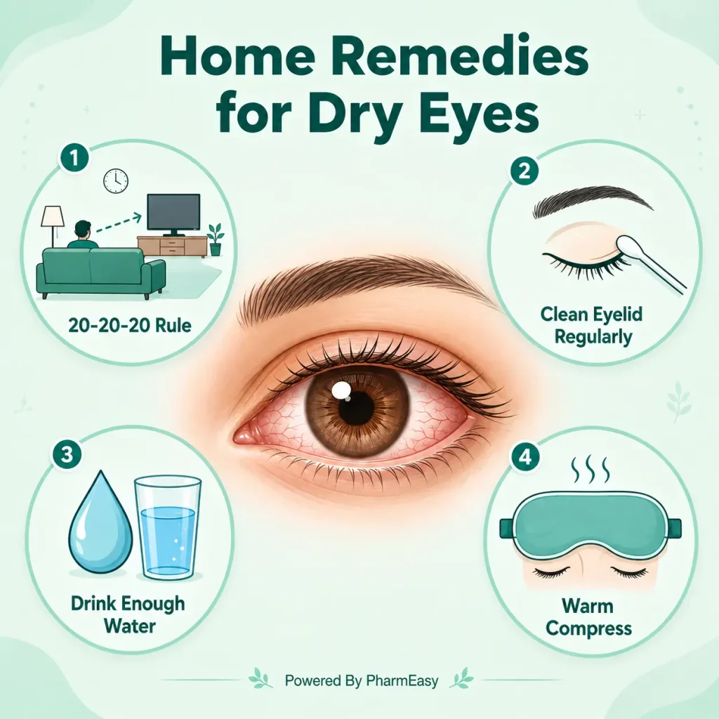
You can manage mild dry eye symptoms at your home. The following are a few easy home remedies for dry eyes.
Note: The information provided is for general awareness only and is not a substitute for professional medical advice, diagnosis, or treatment. If you have persistent or worsening dry eye symptoms, please consult an eye care professional for proper evaluation and management.
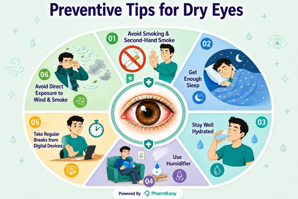
The following are a few ways by which you can avoid having dry eyes:
If your dry eye symptoms persist, worsen, or interfere with your regular activities, you should see an eye doctor. Seek medical treatment if you have severe eye pain, redness, light sensitivity, discharge, or unexpected changes in vision. Early diagnosis and treatment can assist to avoid problems and enhance long-term eye comfort4.
Dry eye syndrome is a common condition that may affect comfort, vision, and overall well-being. It can develop because of ageing, environmental factors, medical issues, or excessive screen time. Understanding the causes, symptoms, and treatment choices will help you manage the disease more effectively. Most people can successfully lessen symptoms and maintain healthy, comfortable eyes by following proper eye care, making lifestyle changes, and seeking timely medical guidance.
Also Read: Sunken Eyes: Causes, Symptoms, Home Remedies & More
You can manage dry eye by using lubricating eye drops, taking regular screen breaks (like the 20-20-20 rule), and avoiding dry or windy environments. Warm compresses and good eyelid hygiene can also help improve tear quality. If symptoms continue, it’s best to consult an eye doctor for proper treatment2,10,15,16.
Dry eye can often be improved and well controlled with treatment during the initial stages. But it is not always completely reversible, especially if it is caused by long-term or chronic conditions. Early diagnosis and proper care can significantly reduce symptoms and prevent it from getting worse1.
Severe dry eye is an advanced form of dry eye where the eyes have very little tear production or significant damage to the eye surface. It can cause scarring of the eye’s surface or damage to the cornea9.
Vitamins A, D, and E, along with omega-3 fatty acids, may help support tear production and maintain eye health17. A balanced diet rich in these nutrients can help reduce dry eye symptoms in some people.
Dry eye can be a long-term condition for some people, especially when linked to ageing or underlying health conditions. However, symptoms can often be effectively managed with proper treatment and lifestyle changes16.
1. Aragona P, Barabino S, Di Zazzo A, et al. Dry Eye Disease: From Causes to Patient Care and Clinical Collaboration—A Narrative Review. Ophthalmol Ther. 2025;14(7):1411-1428. doi:10.1007/s40123-025-01161-8. Available from: https://pubmed.ncbi.nlm.nih.gov/40434534/
2. Better Health Channel. Dry eye [Internet]. 2023 Nov 8 [cited 2026 Jun 12]. Available from: https://www.betterhealth.vic.gov.au/health/conditionsandtreatments/dry-eye
3. National Eye Institute. How tears work [Internet]. Bethesda (MD): National Eye Institute, National Institutes of Health; 2025 Aug 6 [cited 2026 Jun 12]. Available from: https://www.nei.nih.gov/eye-health-information/healthy-vision/how-eyes-work/how-tears-work
4. Healthdirect Australia. Dry eyes [Internet]. Sydney (AU): Healthdirect Australia; reviewed 2024 May [cited 2026 Jun 12]. Available from: https://www.healthdirect.gov.au/dry-eye-disease
5. American Academy of Ophthalmology. Facts about tears [Internet]. San Francisco (CA): American Academy of Ophthalmology; 2024 Mar 15 [cited 2026 Jun 12]. Available from: https://www.aao.org/eye-health/tips-prevention/facts-about-tears
6. Ortega J, Plaska CR, Gomes BA, Ellmore TM. Spontaneous Eye Blink Rate During the Working Memory Delay Period Predicts Task Accuracy. Front Psychol. 2022;13:788231. doi:10.3389/fpsyg.2022.788231. Available from: https://www.frontiersin.org/journals/psychology/articles/10.3389/fpsyg.2022.788231/full
7. American Academy of Ophthalmology. How hormones can affect eyes and vision [Internet]. San Francisco (CA): American Academy of Ophthalmology; 2024 Jun 18 [cited 2026 Jun 12]. Available from: https://www.aao.org/eye-health/tips-prevention/how-hormones-can-affect-eyes-vision
8. Waghmare SV, Jeria S. A Review of Contact Lens-Related Risk Factors and Complications. Cureus. Published online October 10, 2022. doi:10.7759/cureus.30118. Available from: https://pubmed.ncbi.nlm.nih.gov/36381898/
9. Messmer EM. The Pathophysiology, Diagnosis, and Treatment of Dry Eye Disease. Deutsches Ärzteblatt international. Published online January 30, 2015. doi:10.3238/arztebl.2015.0071. Available from: https://pubmed.ncbi.nlm.nih.gov/25686388/
10. Narang P, Donthineni PR, D’Souza S, Basu S. Evaporative dry eye disease due to meibomian gland dysfunction: Preferred practice pattern guidelines for diagnosis and treatment. Indian Journal of Ophthalmology. 2023;71(4):1348-1356. doi:10.4103/IJO.IJO_2841_22. Available from: https://pubmed.ncbi.nlm.nih.gov/37026266/
11. Donthineni PR, Doctor MB, Shanbhag S, et al. Aqueous-deficient dry eye disease: Preferred practice pattern guidelines on clinical approach, diagnosis, and management. Indian Journal of Ophthalmology. 2023;71(4):1332-1347. doi:10.4103/IJO.IJO_2808_22. Available from: https://pubmed.ncbi.nlm.nih.gov/37026265/
12. American Academy of Ophthalmology. Lubricating eye drops for dry eyes [Internet]. San Francisco (CA): American Academy of Ophthalmology; 2024 Sep 10 [cited 2026 Jun 12]. Available from: https://www.aao.org/eye-health/treatments/lubricating-eye-drops
13. De Oliveira RC, Wilson SE. Practical guidance for the use of cyclosporine ophthalmic solutions in the management of dry eye disease. OPTH. 2019;Volume 13:1115-1122. doi:10.2147/OPTH.S184412. Available from: https://pmc.ncbi.nlm.nih.gov/articles/PMC6612764/
14. Rafizadeh SM, Asadigandomani H, Khannejad S, et al. Oculoplastic Interventions in the Management of Ocular Surface Diseases: A Comprehensive Review. Life. 2025;15(7):1110. doi:10.3390/life15071110. Available from: https://pubmed.ncbi.nlm.nih.gov/40724611/
15. Findlay Q, Reid K. Dry eye disease: when to treat and when to refer. Aust Prescr. 2018;41(5):160-163. doi:10.18773/austprescr.2018.048. Available from: https://pubmed.ncbi.nlm.nih.gov/30410213/
16. National Eye Institute. Dry eye [Internet]. Bethesda (MD): National Eye Institute, National Institutes of Health; 2025 Aug 6 [cited 2026 Jun 12]. Available fromi: https://www.nei.nih.gov/eye-health-information/eye-conditions-and-diseases/dry-eye
17. Cong Y, Zhang Y, Han Y, Wu Y, Wang D, Zhang B. Recommendations for nutritional supplements for dry eye disease: current advances. Front Pharmacol. 2024;15:1388787. doi:10.3389/fphar.2024.1388787. Also Read: https://pubmed.ncbi.nlm.nih.gov/38873421/
Disclaimer: The information provided here is for educational/awareness purposes only and is not intended to be a substitute for medical treatment by a healthcare professional and should not be relied upon to diagnose or treat any medical condition. The reader should consult a registered medical practitioner to determine the appropriateness of the information and before consuming any medication. PharmEasy does not provide any guarantee or warranty (express or implied) regarding the accuracy, adequacy, completeness, legality, reliability or usefulness of the information; and disclaims any liability arising thereof.
Links and product recommendations in the information provided here are advertisements of third-party products available on the website. PharmEasy does not make any representation on the accuracy or suitability of such products/services. Advertisements do not influence the editorial decisions or content. The information in this blog is subject to change without notice. The authors and administrators reserve the right to modify, add, or remove content without notification. It is your responsibility to review this disclaimer regularly for any changes.