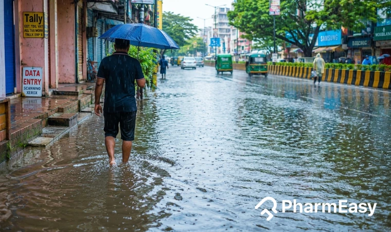
PharmEasy Blog

Protein in Urine (Proteinuria): What Is It, Causes, Symptoms, Diagnosis & More

Introduction

Protein in urine, or proteinuria, is a common condition that can affect people of any age. Sometimes, it is temporary and not serious. However, if it persists or occurs in large amounts, it may indicate an underlying health problem, especially related to the kidneys. The kidneys normally filter waste while keeping important substances like proteins in the blood. When they are not working properly, proteins can leak into the urine. This makes proteinuria an early sign of possible kidney disease1.

Early diagnosis is very important because it can help detect kidney problems or other health conditions at an early stage1.

This article can guide to better understand what proteinuria is, why it happens, its causes and symptoms, how it is diagnosed, and the available treatment options.

What is Proteinuria?

Proteins are important substances that are essential to keep your body healthy. They help give you energy, build and repair muscles, and support your immune system. They can be seen throughout the body, including your blood1.

Your kidneys work like natural filters, cleaning your blood and getting rid of toxins. They keep important substances like proteins in your body while removing waste and extra fluids as urine. This filtering process is very careful, so useful things are not lost.

When the kidneys are affected by disease or when these filters get damaged, they may not work properly. As a result, proteins that should stay in the blood can leak into the urine. This condition is called proteinuria1.

The leakage of protein into the urine can also be due to other reasons like infections, high blood pressure (BP), diabetes, or even temporary conditions like stress, fever, or intense physical activity2. In some cases, proteinuria associated conditions may not cause any noticeable symptoms, especially in the early stages, which is why testing is important for detection1.

Reasons for Protein in Urine

Proteinuria can occur due to several reasons. The following are some of the most common proteinuria causes.

Protein in Urine Symptoms

Protein in urine often does not cause symptoms in the early stages. When symptoms do appear, they may indicate kidney problem or malnutrition caused by protein loss. These symptoms may include:

Types of Proteinuria

Based on the duration, proteinuria can be classified into three

1. Transient Proteinuria

Transient proteinuria is a temporary condition. The protein is found in the urine for a short time, but it goes away once the cause is fixed or disappears. It usually does not mean there is permanent kidney damage. This can happen during short-term situations like fever, seizures, or after heavy exercise, when the body is under stress. It may also occur due to emotional stress, dehydration8, exposure to cold weather or pregnancy2. Sometimes, no obvious reason is found8.

2. Orthostatic Proteinuria

Orthostatic proteinuria is a common condition in children and teenagers, especially boys during adolescence. In this, protein appears in the urine only when the person has been standing or active for several hours. The exact cause is not fully known, but it may be related to changes in blood flow

in the left kidney due to slight pressure on a kidney vein when standing. It is usually a harmless condition and does not indicate any significant health problems8.

3. Persistent Proteinuria

Persistent proteinuria indicates the consistent presence of protein in urine. It usually indicates a kidney problem, most often involving the kidney’s filtering units (glomeruli). When these filters are damaged, proteins like albumin can leak into the urine8. Persistent proteinuria is associated with conditions such as glomerulonephritis, amyloidosis, hypertension, autoimmune disorders, diabetic nephropathy, myeloma, etc2.

How is Protein in Urine Diagnosed?

If you have symptoms associated with proteinuria, your doctor might recommend the following tests for you based on your condition:

Category24-hour Urine ProteinProtein/Creatinine Ratio (mmol)
NormalLess than 150 mg/24 hoursLess than 15 mg/mmol
Mild-Moderate (Nephritic)150-3000 mg/24 hours12-300 mg/mmol
Severe (Nephrotic)More than 3500 mg/24 hoursMore than 350 mg/mmol

The table shows three levels of protein in urine based on how much protein is lost. Normal means only a small, healthy amount of protein is present. The nephritic range means there is a mild to moderate increase, which may suggest early kidney problems or inflammation. The nephrotic range means a very high protein loss, usually indicating serious kidney disease that needs medical care2.

Protein in Urine Treatment

The following are some common treatment strategies used to deal with proteinuria:

Note: Not all medicines are needed for everyone. Treatment varies from person to person and will be prescribed by your doctor based on your condition. Medicines should only be taken under the guidance of your doctor. Self-medication can harm your health or may not provide the expected effectiveness, if not taken with guidance.

How to Reduce Protein in Urine Naturally?

protein in urine

The following are some ways by which you may avoid protein loss:

Note: These suggestions are for general informational purposes only and are not a substitute for medical advice. Protein in urine may indicate an underlying health condition, so proper diagnosis and treatment should be done by a qualified healthcare professional. Always consult your doctor before making any dietary or lifestyle changes.

Also Read: Mucus in Urine: Is It Normal or a Sign of Infection?

When to Consult a Doctor?

You should consult a doctor if protein in urine is detected in a test or if you notice symptoms that may indicate kidney problems. Early medical evaluation is important to prevent complications and identify the underlying cause.

Seek medical attention if you notice:

Even if there are no symptoms, repeated detection of protein in urine in routine tests should always be evaluated by a doctor, as it may be an early sign of kidney disease.

Conclusion

Protein in urine (proteinuria) is often an early sign that the kidneys may not be working properly. It can also occur due to infections, dehydration, or during pregnancy. In many cases, it is temporary, but persistent protein loss should not be ignored. Early testing and treatment can help protect kidney health and avoid serious complications. If you notice symptoms or abnormal test results, it is important to consult a doctor for proper evaluation.

Also Read: Crystals in Urine: Types, Causes, Symptoms & When to Worry

Frequently Asked Questions (FAQs)

What food causes protein in urine?

Reasons for protein in urine can be many and certain types of diet can stress the kidneys or aggravate existing medical issues that can affect the kidneys. Eating too much red meat, processed foods like bacon and sausage, and foods high in salt or sugar can worsen kidney disease, which is a major cause of proteinuria2,3.

What does it mean when you have protein in your urine while pregnant?

Protein in urine during pregnancy is quite common. It can sometimes be harmless and temporary, but in other cases, it may indicate a more serious health problem such as preeclampsia (pregnancy condition marked by high blood pressure and protein in the urine, which can affect both the mother and baby if not treated) that needs medical attention21.

Can alcohol cause protein in urine?

Yes. Frequent consumption of high amounts of alcohol can cause proteinuria7.

Will drinking water reduce protein in urine?

Drinking water can reduce dehydration and help maintain fluid balance in your body by supporting kidney health17, thereby lowering the chances of protein loss. However, it may not stop protein loss caused by severe conditions like CKD.

Can protein in urine go back to normal?

Yes, protein in urine (proteinuria) can return to normal, depending on the cause. It is often temporary and may go away if it is caused by factors like dehydration, heavy exercise, stress, or fever2.

Can UTI cause protein in urine?

Yes, UTIs are known to cause temporary or transient protein loss2. However, it should not be ignored as it can even affect the kidney health and cause severe kidney problems if left untreated. A protein in urine test may help you know its severity.

References

  1. Protein in Urine. 2024. doi: https://medlineplus.gov/lab-tests/protein-in-urine/
  2. Haider MZ, Aslam A. Proteinuria. 2023. doi: https://www.ncbi.nlm.nih.gov/books/NBK564390/
  3. High Blood Pressure & Kidney Disease. 2020. doi: https://www.niddk.nih.gov/health-information/kidney-disease/high-blood-pressure
  4. Lupus & Kidney Disease (Lupus Nephritis). 2017. doi: https://www.niddk.nih.gov/health-information/kidney-disease/lupus-nephritis
  5. Myeloma. doi: https://www.hematology.org/education/patients/blood-cancers/myeloma
  6. Bustamante JG, Zaidi SRH. Amyloidosis. 2023. doi: https://www.ncbi.nlm.nih.gov/books/NBK470285/
  7. Uehara S, Hayashi T, Kogawa Sato K, et al. Relationship Between Alcohol Drinking Pattern and Risk of Proteinuria: The Kansai Healthcare Study. J Epidemiol. 2016;26(9):464-470. doi:10.2188/jea.JE20150158. https://www.jstage.jst.go.jp/article/jea/26/9/26_JE20150158/_article
  8. Leung AKC, Wong AHC, Barg SSN. Proteinuria in Children: Evaluation and Differential Diagnosis. Am Fam Physician. 2017;95(4):248-254. doi: https://www.aafp.org/pubs/afp/issues/2017/0215/p248.html
  9. Understanding your lab values and other CKD health numbers. 2023. doi: https://www.kidney.org/kidney-topics/understanding-your-lab-values-and-other-ckd-health-numbers
  10. Protein electrophoresis – serum. 2024. doi: https://medlineplus.gov/ency/article/003540.htm
  11. ANA (Antinuclear Antibody) Test. 2025. doi: https://medlineplus.gov/lab-tests/ana-antinuclear-antibody-test/
  12. Busuioc R, Ștefan G, Stancu S, Zugravu A, Mircescu G. Nephrotic Syndrome and Statin Therapy: An Outcome Analysis. Medicina. 2023;59(3):512. doi:10.3390/medicina59030512. https://www.mdpi.com/1648-9144/59/3/512/notes
  13. Kang SC, Kim HW, Chang TI, et al. Reduction in proteinuria after immunosuppressive therapy and long-term kidney outcomes in patients with immunoglobulin A nephropathy. Korean J Intern Med. 2021;36(5):1169-1180. doi:10.3904/kjim.2020.240. https://www.kjim.org/upload/kjim-2020-240.pdf
  14. Aggarwal N, Leslie SW. Recurrent Urinary Tract Infections. 2025. doi: https://www.ncbi.nlm.nih.gov/books/NBK557479/
  15. Swift PA, Markandu ND, Sagnella GA, He FJ, MacGregor GA. Modest Salt Reduction Reduces Blood Pressure and Urine Protein Excretion in Black Hypertensives: A Randomized Control Trial. Hypertension. 2005;46(2):308-312. doi:10.1161/01.HYP.0000172662.12480.7f. https://pubmed.ncbi.nlm.nih.gov/15983240/
  16. Michail A, Andreou E. A Plant-Dominant Low-Protein Diet in Chronic Kidney Disease Management: A Narrative Review with Considerations for Cyprus. Nutrients. 2025;17(6):970. doi:10.3390/nu17060970. https://www.mdpi.com/2072-6643/17/6/970
  17. Healthy Hydration and Your Kidneys. 2024. doi: https://www.kidney.org/kidney-topics/healthy-hydration-and-your-kidneys
  18. Managing Diabetes. doi: https://www.niddk.nih.gov/health-information/diabetes/overview/managing-diabetes
  19. Rahaman MdM, Hossain R, Herrera‐Bravo J, et al. Natural antioxidants from some fruits, seeds, foods, natural products, and associated health benefits: An update. Food Science & Nutrition. 2023;11(4):1657-1670. doi:10.1002/fsn3.3217. https://pmc.ncbi.nlm.nih.gov/articles/PMC10084981/
  20. Diabetes Meal Planning. 2024. doi: https://www.cdc.gov/diabetes/healthy-eating/diabetes-meal-planning.html
  21. Airoldi J, Weinstein L. Clinical Significance of Proteinuria in Pregnancy: Obstetrical & Gynecological Survey. 2007;62(2):117-124. doi:10.1097/01.ogx.0000253301.55009.ac. https://pubmed.ncbi.nlm.nih.gov/17229328/

Disclaimer: The information provided here is for educational/awareness purposes only and is not intended to be a substitute for medical treatment by a healthcare professional and should not be relied upon to diagnose or treat any medical condition. The reader should consult a registered medical practitioner to determine the appropriateness of the information and before consuming any medication. PharmEasy does not provide any guarantee or warranty (express or implied) regarding the accuracy, adequacy, completeness, legality, reliability or usefulness of the information and disclaims any liability arising thereof.

Links and product recommendations in the information provided here are advertisements of third-party products available on the website. PharmEasy does not make any representation on the accuracy or suitability of such products/services. Advertisements do not influence the editorial decisions or content. The information in this blog is subject to change without notice. The authors and administrators reserve the right to modify, add, or remove content without notification. It is your responsibility to review this disclaimer regularly for any changes.

Likes
Dislikes

Crystals in Urine: Types, Causes, Symptoms & When to Worry

Introduction

Every day, your body removes waste from the blood through urine. During this process, different minerals and substances dissolve in urine and are excreted along with it. Sometimes, these can stick together and form tiny solid particles called crystals1.

Crystals in urine are very common and are found using routine urine tests. In most cases, a small amount of these crystals is completely normal and may not cause much problems. However, sometimes these crystals can be a sign of a problem. When they appear in large amounts or are of certain types, they may be linked to conditions like kidney stones, dehydration2, urinary tract infections, or metabolic disorders. If not noticed, they can grow or lead to complications1.

In this article, we will discuss the different types of urine crystals, their causes, symptoms, and when to worry. We will also cover how they are diagnosed and how to manage and prevent them.

What Are Crystals in Urine?

Your urine normally contains waste substances like minerals, salts, and acids that are dissolved in liquid. These substances are filtered out of the blood by the kidneys and carried out of the body in urine. Most of the time, they remain dissolved and pass out easily. However, sometimes these substances stick together and form tiny solid particles called crystals. These crystals are usually formed in your kidneys1.

Urine crystals occur when the natural balance of chemicals in urine is disrupted. This can happen if you don’t drink enough water, which makes your urine more concentrated and allows particles to bind together more easily. Crystals can also form when the urine contains too many minerals or compounds. Changes in urine acidity, whether too acidic or alkaline, can also raise the risk1,2.

When this happens, the substances start sticking together and form crystals. Over time, they can grow bigger and may lead to problems like kidney stones.

Causes of Crystals in Urine

Crystals in urine can form due to several reasons that affect the balance of substances in the urine. These include the following.

Crystals in Urine Symptoms

Crystals in urine are often asymptomatic (no symptoms). But when symptoms occur, they may be associated with conditions such as kidney stones, urinary tract infections (UTIs), etc. The following are some symptoms that should not be ignored:

Types of Crystals in Urine

There are several types of urine crystals. The following are the most common types of crystals in urine.

1. Calcium Oxalate Crystals

Calcium oxalate crystals usually contain calcium oxalate, sometimes mixed with calcium phosphate. They are colourless, and shapes can be oval, biconvex, dumbbell-shaped, or long and rod-like may indicate a possible risk of kidney stone formation.

They may also be found in normal urine in small amounts without any disease.

2. Calcium Phosphate Crystals

Contain crystals made of different forms of calcium phosphate such as calcium orthophosphate (amorphous phosphate), carbapatite, and brushite (dicalcium phosphate dihydrate). These crystals are usually colourless and may appear in different shapes depending on the type and may indicate a possible risk of kidney stone formation.

In some cases, they may also be found in normal urine in small amounts without any disease.

3. Struvite (Triple Phosphate Crystals)

Crystals made of magnesium, ammonium, and phosphate. These are colourless and most commonly look like a coffin lid but can also appear as feather-like shapes, long prisms, and trapezoids.

4. Uric Acid Crystals

These include crystals made from four types of uric acid: amorphous uric acid, anhydrous uric acid (uricite), monohydrate, and dihydrate crystals. Among these, amorphous uric acid and uric acid dihydrate are most commonly seen.

They may also appear even when blood uric acid levels are normal.

5. Other Rare Crystals

Some crystals are less common and are usually linked to specific genetic or metabolic conditions. These include:

How Are Crystals in Urine Diagnosed?

Crystals in urine are diagnosed using a combination of several tests, which help identify the type of crystals present and any underlying cause.

Treatment Options for Crystals in Urine

A doctor should always guide treatment for crystals in urine. Hence, it is important to consult a doctor for proper care. He might suggest the following treatment options for crystals in urine.

General Measures

Your doctor might recommend the following measures along with medications:

Medical Treatments

These include the following:

Treating Underlying Conditions: Your doctor suggests treating underlying conditions such as hyperparathyroidism, gout, diabetes, or other metabolic disorders because these conditions can increase certain substances in urine that lead to crystal formation2.

How to Prevent Crystals in Urine?

crystal in urine

The following are some ways by which you can reduce the occurrence of crystals in urine.

When to Consult a Doctor?

The following are symptoms you should not ignore if you have crystals in urine or if you develop any new symptoms.

Conclusion

Crystals in urine are common and often harmless when present in small amounts. However, in some cases, they may indicate dehydration, dietary issues, infections, or underlying medical conditions. Early detection through urine tests helps in proper diagnosis and management. With the right treatment, lifestyle changes, and good hydration, most cases can be effectively managed and avoided.

Also Read: The Fatty Liver Diet: Foods to Eat and Avoid to Reverse Fatty Liver Disease

Frequently Asked Questions (FAQs)

Can a UTI cause crystals in urine?

Yes, a urinary tract infection (UTI) can cause crystals in urine, especially struvite crystals. This happens because certain bacteria change the urine environment, making it more likely for crystals to form2.

What are calcium oxalate crystals in urine?

Calcium oxalate crystals in urine are tiny solid particles made from calcium and oxalate that can form when these substances become too concentrated in urine. They are the most common type of urinary crystals and may sometimes indicate a risk of kidney stones, but they can also appear in healthy people, especially if they are dehydrated or eat foods high in oxalate2.

What to drink for crystals in urine?

Water is the best drink because it helps keep urine diluted and reduces the chance of crystal formation. You may also drink ginger ale or lemon-lime soda to maintain fluid intake11 but avoid too much sugary drinks as they can be dehydrating in summers.

Do urine crystals go away?

Yes, small urine crystals can go away from your body through urine, with low or no pain at all1. However, large crystals might need further medical treatments10.

What foods should you avoid if you have crystals in your urine?

If you are already diagnosed with crystals in urine, you are recommended to avoid foods rich in oxalate (nuts, peanuts, spinach, etc.), animal protein and uric acid (beef, chicken, eggs, shellfish, etc.), and salts (canned, packaged, and fast foods)3.

Are crystals in your urine bad?

Crystals in urine (crystalluria) are not always harmful. While some might (small ones) go away on their own, some can indicate a health problem. They form when there are too many minerals and not enough water in the urine, causing the minerals to stick together1.

References

  1. Crystals in Urine. 2024. doi: https://medlineplus.gov/lab-tests/crystals-in-urine/
  2. Urinary Crystals Identification and Analysis. 2014. doi: https://www.ncbi.nlm.nih.gov/books/NBK606103/
  3. Eating, Diet, & Nutrition for Kidney Stones. doi: https://www.niddk.nih.gov/health-information/urologic-diseases/kidney-stones/eating-diet-nutrition
  4. Kidney Stones. 2024. doi: https://medlineplus.gov/ency/article/000458.htm
  5. Bianchi L, Gaiani F, Bizzarri B, et al. Renal lithiasis and inflammatory bowel diseases, an update on pediatric population. Acta Bio Medica Atenei Parmensis. 2018;89(9-S):76-80. doi:10.23750/abm.v89i9-S.7908: https://pubmed.ncbi.nlm.nih.gov/30561398/
  6. Jalal L, Jaffery Z, Nasir A, et al. Exploring the pathophysiological link between obesity and kidney stone formation: cost-effective dietary and lifestyle strategies for prevention. Annals of Medicine & Surgery. 2025;87(10):6521-6527. doi:10.1097/MS9.0000000000003691: https://pubmed.ncbi.nlm.nih.gov/41181498/
  7. Leslie SW, Sajjad H, Bashir K. 24-Hour Urine Testing for Nephrolithiasis: Interpretation and Treatment Guidelines. 2024. doi: https://www.ncbi.nlm.nih.gov/books/NBK482448/
  8. Urinary Tract Imaging. doi: https://www.niddk.nih.gov/health-information/diagnostic-tests/urinary-tract-imaging
  9. Healthy Hydration and Your Kidneys. 2024. doi: https://www.kidney.org/kidney-topics/healthy-hydration-and-your-kidneys
  10. Treatment for Kidney Stones. doi: https://www.niddk.nih.gov/health-information/urologic-diseases/kidney-stones/treatment
  11. Kidney stones – self-care. 2024. doi: https://medlineplus.gov/ency/patientinstructions/000135.htm

Disclaimer: The information provided here is for educational/awareness purposes only and is not intended to be a substitute for medical treatment by a healthcare professional and should not be relied upon to diagnose or treat any medical condition. The reader should consult a registered medical practitioner to determine the appropriateness of the information and before consuming any medication. PharmEasy does not provide any guarantee or warranty (express or implied) regarding the accuracy, adequacy, completeness, legality, reliability or usefulness of the information; and disclaims any liability arising thereof.

Links and product recommendations in the information provided here are advertisements of third-party products available on the website. PharmEasy does not make any representation on the accuracy or suitability of such products/services. Advertisements do not influence the editorial decisions or content. The information in this blog is subject to change without notice. The authors and administrators reserve the right to modify, add, or remove content without notification. It is your responsibility to review this disclaimer regularly for any changes.

Likes
Dislikes

Bilateral PCOD: Causes, Symptoms, Diagnosis & Treatment

Introduction

Polycystic ovarian changes commonly show up during ultrasound scans in women of reproductive age. They happen when the follicles in the ovaries fail to develop properly and are seen as small fluid-filled sacs on ultrasound scan1,2.  

Polycystic Ovarian Disease (PCOD) or Polycystic Ovarian Syndrome (PCOS) are conditions linked to hormonal imbalances, mainly affecting the ovaries, but can also affect other parts of the body1. Although the terms PCOD and PCOS are commonly used interchangeably, PCOD is a radiographic finding (based on ultrasound scans), while PCOS is the clinically accepted terminology which usually indicates a more severe condition associated with greater hormonal and metabolic disturbances.  

While PCOD and bilateral PCOD seem similar, there is a small difference here. Bilateral PCOD means that both the ovaries show polycystic changes, and unilateral PCOD is when only one ovary is affected3.  

In this blog, we will explain what bilateral PCOD means and how it develops in the body. We will also cover its symptoms, diagnostic methods, and treatment options that can help manage the condition and support better reproductive health. 

What Is Bilateral PCOD?

Polycystic ovary is diagnosed when an ovary has 12 or more small follicles or appears larger than normal (more than 10 cm³) in size as seen on an ultrasound scan. These changes can be seen in one ovary or in both the ovaries4. Bilateral PCOD is a term used when polycystic changes are seen in both the ovaries in an ultrasound scan. The word “bilateral” means “both sides”, so it indicates that changes are present in both the ovaries. Therefore, bilateral PCOD is not a separate disease entity but a way of reporting polycystic changes seen in both the ovaries radiographically3

Although known as polycystic ovaries, the term ‘cyst’ is misleading as these are not actual cysts but small ovarian follicles that fail to develop. Normally, the follicles in the ovary grow and release an egg during ovulation. But in polycystic ovaries, hormone imbalance stops them from maturing properly, so the ovary may have many small, underdeveloped follicles5.  

In bilateral polycystic ovaries, both ovaries may show similar structural changes such as: 

As the follicles don’t develop properly, egg may not be released regularly from the ovaries, affecting the normal menstrual cycle and may even impact fertility, depending on the severity of the condition1,6

Causes of Bilateral PCOD

PCOD or PCOS can develop due to a combination of hormonal, genetic, metabolic, lifestyle, and inflammatory factors that together disrupt normal ovarian function. 

Symptoms of Bilateral PCOD

Symptoms of Bilateral PCOD may range from being asymptomatic (no symptoms, only ultrasound findings) to having severe symptoms similar to PCOS, including: 

Diagnosis of Bilateral PCOD

Bilateral PCOD is a radiographic term (based on ultrasound scan) and does not have a definite diagnostic criterion as per standard guidelines. However, generally along with an ultrasound, your doctor will take a proper medical history and may suggest some blood tests to understand the impact of the condition. 

1. Medical History

Your doctor will discuss your symptoms and do an overall assessment. They may ask for: 

2. Hormonal Blood Test

Your doctor might suggest several blood tests to understand whether you have PCOD or are at a risk of developing it1. She might suggest tests such as: 

3. Ultrasound

Ultrasound findings are important to diagnose Bilateral PCOD. An Ultrasound scan will show multiple follicles in the ovaries through high-frequency sound waves and produces clear images. Their types include: 

Note: The diagnosis of PCOS is based on the Rotterdam criteria7 and requires the presence of least 2 of the following 3, irrespective of unilateral or bilateral involvement of ovaries: Oligo or Anovulation (egg may not mature properly or may fail to be released during ovulation), Hyperandrogenism (high levels of male sex hormones), Polycystic Ovarian Changes (seen on ultrasound).  

Treatment of Bilateral PCOD

Treatment for polycystic ovaries focuses on managing symptoms, improving fertility, and avoiding long-term problems. While there is no permanent solution, treatment is personalised to help achieve goals like regular periods, clearer skin, reduced hair growth, and improved chances of pregnancy. 

Treatment options that help manage PCOD and its symptoms: 

Treatment options if you want to get pregnant while having polycystic ovaries: 

Note: All the tests and treatments mentioned in this section should only be carried out under the guidance of a qualified gynaecologist and cosmetologist. Self-medication is not recommended. Ignoring professional advice and taking over-the-counter medicines may not provide benefits and can also negatively affect your health. 

Home Remedies for Bilateral PCOD

home remedies for bilateral pcod

The following are some home remedies that may help in managing polycystic ovaries symptoms and improving overall hormonal balance. 

Note: Although home care measures can help manage the symptoms of polycystic ovaries, they should not replace proper medical treatment. Always consult your doctor before attempting weight loss or using herbal supplements. These remedies should be used alongside prescribed treatment, not as a substitute. Consult a doctor if the symptoms get worse. 

Also Read: PCOD Diet Chart: Foods to Eat, Avoid, Meal Plan & Lifestyle Tips 

When to Consult a Doctor?

The following are some symptoms you should not ignore, whether or not you have bilateral PCOD. 

If you have already been diagnosed with bilateral PCOD, it is important to regularly monitor your symptoms. Even if you are not diagnosed but experience similar symptoms, consult your doctor for proper evaluation and guidance. 

Also Read: PCOS Diet: How to Use Food to Help Manage Your PCOS

Conclusion

Now you might be clear with the bilateral PCOD meaning; it is caused when both ovaries are affected by changes caused by a hormone imbalance. While it may cause symptoms like irregular periods, weight changes, and skin issues, it can be managed effectively with proper treatment and lifestyle changes. Early diagnosis and regular medical guidance play an important role in avoiding complications. With the right care, many women can maintain good reproductive and overall health. 

Frequently Asked Questions (FAQs)

Is bilateral PCOD serious? 

Bilateral PCOD is usually not serious, but it can cause symptoms like irregular periods, weight gain, and hormonal imbalance. If not managed, it may lead to complications like diabetes or fertility issues1

Can you get pregnant with bilateral PCOD? 

Yes, many women with bilateral PCOD can get pregnant, either naturally or with treatment. Proper management can improve ovulation and increase chances of pregnancy11

Can bilateral PCOD be cured? 

Bilateral PCOD is chronic but manageable condition. But it can be effectively managed with lifestyle changes and medical treatment to manage symptoms and improve overall health1,2

What is the bilateral PCOD pattern? 

The bilateral PCOD pattern indicates that, on ultrasound, both ovaries appear enlarged and contain many small follicles, which are typical PCOD changes1,3,5

At what age does bilateral PCOD occur? 

Bilateral PCOD or PCOS often starts around puberty, sometimes as early as 11–12 years during the first menstrual cycles, but it can also develop later in life1

How often should someone with bilateral PCOD see a doctor? 

Women with polycystic ovaries should have regular follow-ups. High-risk patients (metabolic problems, obesity, or severe symptoms) may need check-ups every 6 months, while low-risk patients (mild or well-controlled symptoms with no major complications) can visit once a year to monitor their health. 

What happens if bilateral PCOD or PCOS is not managed properly? 

Untreated bilateral PCOD/PCOS can lead to serious health problems such as diabetes, heart disease, high blood pressure, sleep disorders, pregnancy complications, and infertility2. It can also increase the risk of endometrial cancer, as lack of ovulation (chronic anovulation) leads to prolonged oestrogen  exposure and thickening of the uterine lining16

What are the common myths about bilateral PCOD or PCOS? 

A common myth is that PCOD or PCOS is caused by ovarian cysts. However, the small follicles seen in PCOS are not the cause of the condition. It is mainly caused by hormonal imbalance. Another misconception is that women with PCOS cannot get pregnant, but many can conceive naturally or with treatment17

References

1. Polycystic Ovary Syndrome. 2024. Doi: https://medlineplus.gov/polycysticovarysyndrome.html 

2. Polycystic ovarian syndrome (PCOS). 2024. Doi: https://www.healthdirect.gov.au/polycystic-ovarian-syndrome-pcos#symptoms 

3. Leelamma J, Pillai MT, S A, Nambisan B, Nambiar R. Comparison between unilateral and bilateral polycystic ovaries in adolescent PCOS. Scholars Journal of Applied Medical Sciences (SJAMS). 5(6):2472-2477. Doi: https://www.saspublishers.com/article/14089/download/ 

4. Christ JP, Cedars MI. Current Guidelines for Diagnosing PCOS. Diagnostics. 2023;13(6):1113. doi:10.3390/diagnostics13061113. https://pubmed.ncbi.nlm.nih.gov/36980421/ 

5. Polycystic ovary syndrome. 2020. Doi: https://medlineplus.gov/genetics/condition/polycystic-ovary-syndrome/ 

6. Nallaparaju LS. QUANTIFYING THE PREVALENCE AND INTERDEPENDENT RELATIONSHIPOF PCOD, OBESITY, AND DEPRESSION – A PROSPECTIVE OBSERVATIONAL, POLYCENTRIC STUDY. World Journal of Pharmaceutical Research. 12(7):732-764. Doi: https://wjpr.s3.ap-south-1.amazonaws.com/article_issue/57fb03a2e4edae203d6b8262b656e58b.pdf 

7. Sadeghi HM, Adeli I, Calina D, et al. Polycystic Ovary Syndrome: A Comprehensive Review of Pathogenesis, Management, and Drug Repurposing. IJMS. 2022;23(2):583. doi:10.3390/ijms23020583. https://pubmed.ncbi.nlm.nih.gov/35054768/ 

8. Anti-Müllerian Hormone Test. 2023. Doi: https://medlineplus.gov/lab-tests/anti-mullerian-hormone-test/ 

9. Prosperi S, Chiarelli F. Insulin resistance, metabolic syndrome and polycystic ovaries: an intriguing conundrum. Front Endocrinol. 2025;16:1669716. doi:10.3389/fendo.2025.1669716. https://www.frontiersin.org/journals/endocrinology/articles/10.3389/fendo.2025.1669716/full 

10. Di Michele S, Fulghesu AM, Pittui E, et al. Ultrasound Assessment in Polycystic Ovary Syndrome Diagnosis: From Origins to Future Perspectives—A Comprehensive Review. Biomedicines. 2025;13(2):453. doi:10.3390/biomedicines13020453. https://pubmed.ncbi.nlm.nih.gov/40002866/ 

11. Polycystic ovary syndrome: OASH. 2025. Doi: https://womenshealth.gov/a-z-topics/polycystic-ovary-syndrome 

12. Paramasivam A, Murugan R, Jeraud M, Dakkumadugula A, Periyasamy R, Arjunan S. Additives in Processed Foods as a Potential Source of Endocrine-Disrupting Chemicals: A Review. JoX. 2024;14(4):1697-1710. doi:10.3390/jox14040090. https://pubmed.ncbi.nlm.nih.gov/39584955/ 

13. Rao V, Pena A, James A, et al. The role of meditation and mindfulness in the management of polycystic ovary syndrome: a scoping review. Front Endocrinol. 2024;15:1295705. doi:10.3389/fendo.2024.1295705. https://pubmed.ncbi.nlm.nih.gov/38818503/ 

14. Goodarzi L, Ahmadi MM, Ramezanirad M, et al. The Role of Sleep Hygiene in Different Patterns of Polycystic Ovary Syndrome (PCOS): Insights from Modern and Persian Medicine. MJIRI. Published online February 25, 2025. doi:10.47176/mjiri.39.116. https://pmc.ncbi.nlm.nih.gov/articles/PMC12584089/ 

15. Muhammed Saeed AA, Noreen S, Awlqadr FH, et al. Nutritional and herbal interventions for polycystic ovary syndrome (PCOS): a comprehensive review of dietary approaches, macronutrient impact, and herbal medicine in management. J Health Popul Nutr. 2025;44(1):143. doi:10.1186/s41043-025-00899-y. https://pubmed.ncbi.nlm.nih.gov/40317096/ 

16. Bassette E, Ducie JA. Endometrial Cancer in Reproductive-Aged Females: Etiology and Pathogenesis. Biomedicines. 2024;12(4):886. doi:10.3390/biomedicines12040886. https://pubmed.ncbi.nlm.nih.gov/38672240/ 

17. Polycystic Ovary Syndrome (PCOS) Blog. 2024. Doi: https://www.fda.gov/consumers/knowledge-and-news-women-owh-blog/polycystic-ovary-syndrome-pcos-blog 

Disclaimer: The information provided here is for educational/awareness purposes only and is not intended to be a substitute for medical treatment by a healthcare professional and should not be relied upon to diagnose or treat any medical condition. The reader should consult a registered medical practitioner to determine the appropriateness of the information and before consuming any medication. PharmEasy does not provide any guarantee or warranty (express or implied) regarding the accuracy, adequacy, completeness, legality, reliability or usefulness of the information; and disclaims any liability arising thereof.

Links and product recommendations in the information provided here are advertisements of third-party products available on the website. PharmEasy does not make any representation on the accuracy or suitability of such products/services. Advertisements do not influence the editorial decisions or content. The information in this blog is subject to change without notice. The authors and administrators reserve the right to modify, add, or remove content without notification. It is your responsibility to review this disclaimer regularly for any changes.

Likes
Dislikes

Types of Cancer: Symptoms, Early Signs, Diagnosis & More 

Introduction

Cancer is one of the major health problems around the world, affecting millions of people every year. Even though there is a big advancement in the medical treatment, many cases are detected very late, making the treatment procedure difficult. This is why knowing cancer is important, especially its early signs and symptoms. Finding cancer early can greatly improve the chances of successful treatment and recovery1

There are different types of cancer, which can affect different body parts, and the early symptoms are often not clear or easy to notice. Common warning signs may include unexplained weight loss, constant tiredness, unusual lumps, unusual sweeting, etc2. Pain is usually a late feature in many cancers, so paying attention to the other changes in the body and consulting a doctor on time is important  for diagnosis and improving treatment outcomes. 

This article might help you understand the main types of cancer, their early warning signs and how they are diagnosed. The aim is to raise awareness, help detect cancers early, and encourage people to take better care of their health.  

What Is Cancer?

Cancer is a disease that happens when normal cells in the body change and start growing uncontrollably, forming a tumour that can spread and harm healthy tissues1. However, all tumours are not cancers. They can be benign or malignant3. Also, not all cancers form solid tumours, such as blood cancers, which have no discrete growth as such4

The spread of cancer cells from one part of the body to another through the blood or lymph system is called metastasis. In addition, cancer also spread directly into nearby tissues (local invasion)3 or within body cavities, such as the abdomen6.  

The risk of cancer increases with age. This is because, over time, the body builds up more risk factors, and the natural repair system of cells becomes less effective as we grow older1. Other risk factors include family history, lifestyle (smoking, alcohol, etc.), viral infections (human papilloma or hepatitis), etc2.   

Cancer develops differently in each person, and not all growths are harmful. With early detection and proper treatment, many cancers can be managed effectively1.

Main Types of Cancer

There are different types of cancers which can develop in different body parts. Most of these cancers have very few known causes4,7,9. The following might help you understand how many types of cancer exist based on the types of cancer cells. 

1. Carcinoma

Carcinoma is a type of cancer that starts in epithelial tissue, which covers the skin and lines the inside of organs and body passages. It can develop in tissues that produce or secrete substances, such as in the breast, lungs, colon, prostate, or bladder, (adenocarcinoma) or in flat, thin cells that form the skin surface and line some internal organs (squamous cell carcinoma)10. The common cause is the accumulation of genetic changes over time, which leads cells to uncontrolled growth of cells. The risk factors that can trigger these genetic alterations including smoking, alcohol consumption, chemical exposure, lack of physical activity, obesity, etc11,12

2. Sarcoma

Sarcoma is a type of cancer that begins in the body’s supportive and connective tissues like bones, muscles, fat, cartilage, and tendons. It is more common in children and in adolescents, compared to carcinoma. However, several other types (soft tissue sarcomas) can also occur in adults. It often appears as a painful lump, especially in the bones, and the tumour usually looks like the normal tissue from where it starts10. It is commonly caused by mutation in the gene due to exposure to radiation or by certain cancer-causing chemicals (carcinogens)9 

3. Leukaemia

Leukaemia is a blood cancer that begins in the bone marrow, the place where blood cells are produced. It disrupts the normal functioning of bone marrow, leading to the reduced production of red blood cells and the platelet count. This can cause problems like fatigue, anaemia, and poor blood clotting10. The risk factors include family history, age and sex, exposure to harmful radiations and chemicals, and viral infections (human T-cell leukaemia virus)4

4. Myeloma

Myeloma (multiple myeloma) is a type of cancer that affects plasma cells, which are white blood cells that help fight infection by making antibodies8. The plasma cells produce certain types of protein (monoclonal (M) protein/paraprotein) which are found in the blood10. Here, abnormal plasma cells grow too much in the bone marrow and replace healthy blood cells, affecting red cells, white cells, and platelets. The risk factors include age, exposure to x-rays or other radiations, people belonging to specific races, etc8.  

5. Lymphoma

Lymphoma is a type of cancer that starts in the lymphatic system, which includes lymph nodes, spleen, tonsils, and thymus. This system helps fight infections by supporting the development and activation of white blood cells. It is a type of blood-related (hematologic) cancer, that is often seen as enlarged lymph nodes or mass-like growths. It can also develop in organs like the stomach, breast, or brain10. People with weak immune systems, those on immune-suppressing medicines, Epstein-Barr virus infection, radiation exposure, and those with a family history are at higher risk of developing lymphoma7.  

Common Types of Cancer & Their Symptoms

There are several types of cancers. Below are some common types and their symptoms. 

1. Breast Cancer

breast cancer

2. Lung Cancer

lung cancer

3. Colorectal Cancer

colorectal cancer

4. Prostate Cancer

prostate cancer

5. Skin Cancer

skin cancer

Early Signs of Cancer

Although different types of cancer show different signs and symptoms, there are some warning signs to watch out for. The following are some early signs of cancer that you should not ignore. 

Why Early Detection Matters?

Early detection of cancer plays a crucial role in improving outcomes and saving lives. The following says why early detection matters and how it is helpful to patients. 

Note: Early detection helps improve outcomes in many cancers like breast, cervical, and colorectal cancer, but it is not equally effective for all cancers. Some, like pancreatic cancer, are often found late, while others (e.g., prostate or thyroid cancer) may be over diagnosed. Early treatment can improve well-being but may still cause side effects, and even advanced cancers can sometimes be managed with good care. 

Blood Test for Early Cancer Detection

The following is a list of blood tests performed in early cancer detection. 

Basic Tests

Advanced Tests

Note: Diagnosis of cancer typically requires histopathological confirmation (biopsy); imaging or basic blood screening tests alone are generally not definitive for most cancers. 

When to Consult a Doctor?

You should consider seeing a doctor if you notice: 

Also Read: Does Masturbating Increase Risk of Prostate Cancer or Vice Versa?

Conclusion

Cancer is a serious condition, but it can often be effectively managed, especially with early detection. Recognising warning signs and going for regular check-ups can greatly improve treatment outcomes. Understanding different types of cancer and their symptoms helps you stay aware and take timely action. Prioritising your health and consulting a doctor when needed can make a life-saving difference. 

Frequently Asked Questions (FAQs)

Can MRI detect all types of cancer? 

An MRI cannot test all types of cancer. It is good to detect cancer caused in the brain, soft tissues, breast and colon12,14,21. However, it has limitations in early diagnosis of some types of cancers like lung cancer21.

How many types of cancer are there? 

There are said to be more than 200 types of cancer which affect the organs, tissues, blood, bone marrow and immune system2

Can you have multiple types of cancer? 

Yes, it is possible for a person to have more than one type of cancer, either at the same time or one after another. This is called multiple primary cancers. Although it is not very common, these cancers start separately in different parts of the body and are not caused by the spread of an existing cancer22

What are the silent signs of cancer? 

The silent signs of cancer include: 
-Unexplained weight loss 
-Lack of appetite 
-New or unexplained pain2 
-New mole or changes in existing mole (skin changes) 
-Unusual bleeding or bruising 
-Changes in bowel movement 
-Long-term cough or hoarseness17 
-Difficulty in breathing or swallowing, chest pain or discomfort, etc11 

What are the common types of cancer in women? 

The most common types of cancer found in women are breast cancer, cervical cancer, ovarian cancer, uterine cancer, oral cancer and colorectal cancer23

What are the common types of cancer in men? 

The most common types of cancer found in men are prostate cancer, colorectal cancer, lung cancer, oral cancer, stomach cancer and oesophageal cancer23

What type of cancer causes weight loss? 

The types of cancers that cause weight loss include colorectal cancer, lung cancer, gastro-oesophageal cancer, pancreatic cancer, liver cancer, ovarian cancer, etc11,14,24

References

1. Cancer. 2025. Doi: https://www.who.int/news-room/fact-sheets/detail/cancer 

2. Cancer: NHS. 2025. Doi: https://www.nhs.uk/conditions/cancer/ 

3. Cancer: Medline. 2025. Doi: https://medlineplus.gov/cancer.html 

4. Leukaemia. 2023. Doi: https://www.healthdirect.gov.au/leukaemia 

5. Benign. 2025. Doi: https://medlineplus.gov/ency/article/002236.htm 

6. Menon G, Santillan VR. Peritoneal Surface Malignancies. 2025. Doi: https://www.ncbi.nlm.nih.gov/books/NBK541114/ 

7. Lymphoma. 2025. Doi: https://www.healthdirect.gov.au/lymphoma 

8. Myeloma Basics. 2025. Doi: https://www.cdc.gov/myeloma/about/index.html 

9. Causes, Risk Factors, and Prevention of Soft Tissue Sarcomas. 2026. Doi: https://www.cancer.org/cancer/types/soft-tissue-sarcoma/causes-risks-prevention.html 

10. Cancer Classification. Doi: https://training.seer.cancer.gov/disease/categories/classification.html 

11. Lung Cancer. 2025. Doi: https://medlineplus.gov/lungcancer.html 

12. Breast Cancer. 2025. Doi: https://medlineplus.gov/breastcancer.html 

13. Colorectal Cancer. 2024. Doi: https://medlineplus.gov/colorectalcancer.html 

14. Bowel cancer (colon and rectal cancer). 2023. Doi: https://www.healthdirect.gov.au/bowel-cancer 

15. Prostate Cancer. 2024. Doi: https://medlineplus.gov/prostatecancer.html 

16. Sathe NC, Zito PM. Skin Cancer. 2025. Doi: https://www.ncbi.nlm.nih.gov/books/NBK441949/ 

17. Symptoms of Cancer. 2019. Doi: https://www.cancer.gov/about-cancer/diagnosis-staging/symptoms 

18. Imai M, Nakamura Y, Yoshino T. Transforming cancer screening: the potential of multi-cancer early detection (MCED) technologies. Int J Clin Oncol. 2025;30(2):180-193. doi:10.1007/s10147-025-02694-5. https://pubmed.ncbi.nlm.nih.gov/39799530/ 

19. Tumor Marker Tests in Common Use. 2023. Doi: https://www.cancer.gov/about-cancer/diagnosis-staging/diagnosis/tumor-markers-list 

20. How Cancer Is Diagnosed. 2023. Doi: https://www.cancer.gov/about-cancer/diagnosis-staging/diagnosis 

21. Sim AJ, Kaza E, Singer L, Rosenberg SA. A review of the role of MRI in diagnosis and treatment of early stage lung cancer. Clinical and Translational Radiation Oncology. 2020;24:16-22. doi:10.1016/j.ctro.2020.06.002. https://pmc.ncbi.nlm.nih.gov/articles/PMC7306507/ 

22. Vogt A, Schmid S, Heinimann K, et al. Multiple primary tumours: challenges and approaches, a review. ESMO Open. 2017;2(2):e000172. doi:10.1136/esmoopen-2017-000172. https://www.esmoopen.com/article/S2059-7029(20)32451-0/fulltext 

23. Kalra K. Common Cancers in India, theirincreasing incidence. Symptomswhich need immediate attention. Doi: https://www.cghs.mohfw.gov.in/CGHSGrievance/FormFlowXACTION?hmode=ftpFileDownload&fileName=23052025114829_Common-Cancers-in-India-their-increasing-incidence-(9-November-2021)-.pdf&folderName=Circular&isGlobal=1 

24. Nicholson BD, Hamilton W, O’Sullivan J, Aveyard P, Hobbs FR. Weight loss as a predictor of cancer in primary care: a systematic review and meta-analysis. Br J Gen Pract. 2018;68(670):e311-e322. doi:10.3399/bjgp18X695801. https://pubmed.ncbi.nlm.nih.gov/29632004/ 

Disclaimer: The information provided here is for educational/awareness purposes only and is not intended to be a substitute for medical treatment by a healthcare professional and should not be relied upon to diagnose or treat any medical condition. The reader should consult a registered medical practitioner to determine the appropriateness of the information and before consuming any medication. PharmEasy does not provide any guarantee or warranty (express or implied) regarding the accuracy, adequacy, completeness, legality, reliability or usefulness of the information; and disclaims any liability arising thereof.

Links and product recommendations in the information provided here are advertisements of third-party products available on the website. PharmEasy does not make any representation on the accuracy or suitability of such products/services. Advertisements do not influence the editorial decisions or content. The information in this blog is subject to change without notice. The authors and administrators reserve the right to modify, add, or remove content without notification. It is your responsibility to review this disclaimer regularly for any changes.

Likes
Dislikes

Muscle Spasm: Meaning, Causes, Symptoms, Treatment & Prevention

Introduction

Have you ever experienced a sudden painful cramp in your muscle that makes it hard to move? This is called a muscle spasm. It occurs when a muscle contracts unexpectedly but does not relax properly1. Muscle spasms are very common and may occur to anybody despite their age. They can occur during exercise, while resting, or even during sleep, like the familiar leg cramps many people experience at night1,2. In most of the cases, these spasms are harmless and may resolve themselves. However, they can cause discomfort or pain and may interrupt daily activities. 

While most of the muscle cramps are harmless, some can indicate certain underlying health conditions1. Understanding the causes, symptoms, and ways to manage muscle spasms can help you manage them better and stay comfortable. This article may help you understand all of this. 

What is a Muscle Spasm?

A muscle spasm is an involuntary contraction of a muscle a muscle suddenly tightens on its own without you trying to control it. Normally, the muscles contract and relax smoothly, which gives a smooth movement. But during a spasm, the affected muscle contracts unexpectedly and does not relax properly. It can cause sudden, tight and intense pain1.

This can happen in a single muscle or a group of muscles. It can last from a few seconds to several minutes3 and may occur once in a while or repeatedly. It is often triggered by factors such as overuse, dehydration, poor circulation, or electrolyte imbalance3

What Does a Muscle Spasm Feel Like?

A muscle spasm can feel different from person to person, but it is usually sudden and noticeable. It often begins without warning and may last for a few seconds to several minutes. The common muscle spasm symptoms include: 

Causes of Muscle Spasm

Muscle spasm can occur due to several reasons. The following are some common muscle spasm causes. 

Types of Muscle Spasms

Muscle spasms can happen in different parts of the body due to reasons like poor posture, exercise, dehydration, or health issues. The feeling may vary depending on the muscle affected. They include the following:

Are Muscle Spasms Serious?

Most muscle spasms are harmless and temporary. They usually happen due to common causes like dehydration, stress, muscle fatigue or overuse and often go away on their own within a few seconds to minutes. In many cases, simple measures such as rest, gentle stretching, hydration, and applying heat or cold may help relieve the discomfort1,3,8.

However, sometimes they can also indicate some underlying conditions, such as nerve disorders, organ-related problems or electrolyte imbalance leading to severe health problems6. Monitor symptoms and frequency and try improving hydration along with other simple techniques that may help relieve muscle spasm1. If all these do not reduce the spasm issues, then it is good to consult a doctor to identify the cause. 

Thus, occasional muscle spasms can be considered normal, but persistent or severe symptoms, and those which interfere with day-to-day activities, could indicate an underlying issue and should be evaluated by a doctor. 

Diagnosis of Muscle Spasm

Muscle spasm has different diagnostic procedures to identify the causes. They include the following. This might help in deciding which muscle spasm treatment to take.

Note: These are just a few examples; there may be more tests prescribed based on the medical history and symptoms. 

Treatment of Muscle Spasm

The cause of muscle spasm can range from mild to severe. Therefore, your doctor would suggest treatment based on the cause. The following are some treatment options which help to understand how to cure muscle spasm. These are for spasms which are frequent, severe, or linked to underlying conditions: 

Note: The treatment methods described in this section are only to be performed under the guidance of a doctor. Avoid buying the medicines over the counter; instead seek medical attention for proper treatment. 

How to Manage Muscle Spasm Naturally?

muscle spasm

The following are some natural ways you may try to manage muscle spasm: 

Note: The above-mentioned methods are for temporary problems only. These alone may not resolve the problem completely. If you think spasm is not reducing and is still getting worse, please consult a doctor as soon as possible, as this can also indicate severe conditions. 

Prevention Tips for Muscle Spasms

The following are some tips that might help avoid muscle spasm: 

When to See a Doctor?

Consult a doctor immediately if you have muscle spasms, which are: 

Conclusion

So, now you might be clear on muscle spasm meaning. They are common and usually harmless, often caused by factors like dehydration, overuse, or electrolyte imbalance. In most cases, they resolve with simple measures such as rest, hydration, and stretching. However, persistent or severe spasms may indicate an underlying health condition and should not be ignored. Early medical evaluation and proper management might help reduce recurrence and improve quality of life.

Also Read: Food Allergies: Symptoms, Types, Diagnosis & Home Remedies

Frequently Asked Questions (FAQs)

Are muscle spasms dangerous?

Muscle spasms are usually harmless, caused as a result of dehydration, overusing of muscles or poor posture. However, sometimes they can also indicate serious conditions such as nerve problems, organ-related conditions, infections, etc1. Therefore, it is important to analyse the symptoms and its frequency for a proper treatment.

What triggers a muscle spasm?

The factors which trigger muscle spasm include dehydration, electrolyte imbalance, nerve compression, overstraining, poor blood circulation, pregnancy, certain medications and certain underlying conditions4.

How long do muscle spasms normally last?

A muscle spasm can last for a few seconds to 10-15 minutes. However, sometimes, it can extend up to several hours1,4.

What to drink to stop muscle spasms?

If the muscle spasm is due to electrolyte imbalance and dehydration, drinking electrolyte-rich fluids may help replenish the body and replace lost electrolytes4. These include coconut water, orange juice, milk, sports drinks, etc4,16.

What can be mistaken for a muscle spasm?

Many conditions, such as muscle twitching as in tetany, movement disorders like dystonia, or continuous muscle stiffness in myotonia or hypothyroidism etc can be mistaken for a muscle spasm9.

What vitamin deficiency causes muscle spasm?

The deficiencies of vitamin B and D are known to cause muscle spasms4.

References

  1. Muscle Cramps. doi: Available from: https://orthoinfo.aaos.org/en/diseases–conditions/muscle-cramps
  2. Blyton F, Chuter V, Burns J. Unknotting night‐time muscle cramp: a survey of patient experience, help‐seeking behaviour and perceived treatment effectiveness. Journal of Foot and Ankle Research. 2012;5(1):7. doi:10.1186/1757-1146-5-7. Available from: https://pmc.ncbi.nlm.nih.gov/articles/PMC3361473/
  3. Muscle Cramps. 2025. doi: Available from: https://medlineplus.gov/musclecramps.html
  4. Goldin J, Sugumar K. Muscle Cramps. 2025. doi: Available from: https://www.ncbi.nlm.nih.gov/books/NBK499895/
  5. Caring for muscle spasticity or spasms. 2024. doi: Available from: https://medlineplus.gov/ency/patientinstructions/000063.htm
  6. J Lespasio M. Muscle Spasm: A Primer. IJCMCR. 2024;44(4). doi:10.46998/IJCMCR.2024.44.001091. Available from: https://ijclinmedcasereports.com/pdf/IJCMCR-RW-01091.pdf
  7. Aches and pains during pregnancy. 2024. doi: Available from: https://medlineplus.gov/ency/patientinstructions/000580.htm
  8. Parasa M, Saheb S, Vemuri N. Cramps and tingling: A diagnostic conundrum. Anesth Essays Res. 2014;8(2):247. doi:10.4103/0259-1162.134524. Available from: https://pmc.ncbi.nlm.nih.gov/articles/PMC4173627/
  9. Stern LZ, Bernick C. Chapter 53: Muscle Cramps. doi: Available from: https://www.ncbi.nlm.nih.gov/books/NBK376/
  10. Torticollis. 2024. doi: Available from: https://www.healthdirect.gov.au/torticollis
  11. Low back pain – acute. 2024. doi: Available from: https://medlineplus.gov/ency/article/007425.htm
  12. Leg Cramps. 2006. Available from: https://www.nhsinform.scot/illnesses-and-conditions/muscle-bone-and-joints/leg-and-foot-problems-and-conditions/leg-cramps/
  13. Abdominal pain. 2024. doi: Available from: https://medlineplus.gov/ency/article/003120.htm
  14. Hand or foot spasms. 2024. doi: Available from: https://medlineplus.gov/ency/article/003194.htm
  15. Capsaicin Topical. 2020. doi: Available from: https://medlineplus.gov/druginfo/meds/a620055.html
  16. Electrolyte Drinks. 2025. doi: Available from: https://nutritionsource.hsph.harvard.edu/electrolyte-drinks/
  17. Relaxation techniques for stress. 2024. doi: Available from: https://medlineplus.gov/ency/patientinstructions/000874.htm
  18. Muscle cramp. 2013. doi: Available from: https://www.betterhealth.vic.gov.au/health/conditionsandtreatments/muscle-cramp

Disclaimer: The information provided here is for educational/awareness purposes only and is not intended to be a substitute for medical treatment by a healthcare professional and should not be relied upon to diagnose or treat any medical condition. The reader should consult a registered medical practitioner to determine the appropriateness of the information and before consuming any medication. PharmEasy does not provide any guarantee or warranty (express or implied) regarding the accuracy, adequacy, completeness, legality, reliability or usefulness of the information and disclaims any liability arising thereof.

Links and product recommendations in the information provided here are advertisements of third-party products available on the website. PharmEasy does not make any representation on the accuracy or suitability of such products/services. Advertisements do not influence the editorial decisions or content. The information in this blog is subject to change without notice. The authors and administrators reserve the right to modify, add, or remove content without notification. It is your responsibility to review this disclaimer regularly for any changes.

Likes
Dislikes

Double Marker vs. Triple Marker Test: Differences, Timing, Results & How They Work Together 

Introduction

The pregnancy period involves several checkups, which are essential to ensure that the baby is growing well and staying healthy. An important part of these check-ups is early pregnancy (prenatal) screening, which helps doctors understand if there are any chances of certain health conditions in the baby early on. These are simple screening tests, which are different from diagnostic procedures1

A type of screening tests employed for this purpose are the marker tests. These are the blood tests which measure some natural components (biomarkers) in the mother’s blood. The levels of these markers can give an idea about the baby’s risk of conditions, for e.g., some genetic conditions like Down’s syndrome2.

These marker tests include the double marker test and the triple marker test. They are similar but done at different stages of pregnancy and check different types of markers3,4.

In this article, we’ll explain the difference between double marker and triple marker tests, when they are performed, what their results say, and how they can be employed together to assess the baby’s health. 

What Is a Double Marker Test?

The double marker test is a pregnancy screening blood test, which helps assess the risk of certain genetic conditions in the baby during early pregnancy. It is usually performed as a part of first-trimester (first 3 months of pregnancy) screening, which helps determine early indications of whether further detailed testing may be needed3.

The test measures two specific markers present in the mother’s blood: 

The result of these markers may help evaluate the placental and baby’s health (abnormal levels of markers may indicate poor or abnormal formation of the placenta)5. This result is then combined with ultrasound scanning tests (especially nuchal translucency (NT)) and the mother’s age to estimate the possible risk3

This test is usually performed between the 10th and 13th weeks +6 days of gestation (pregnancy)6. Abnormal levels (too high or too low) may help indicate increased risk of conditions like: 

However, for confirmation, the doctors might prescribe several other diagnostic tests. 

What Is a Triple Marker Test?

A triple market test is also a pregnancy screening test, but it is usually performed during the second trimester (4th– 6th month) of pregnancy. This test helps assess the risk of several genetic disorders in the baby, by evaluating certain markers in the mother’s blood, along with the mother’s age, during the pregnancy periods. This, in turn, helps evaluate whether further detailed diagnostic testing for specific conditions may be needed or not2,7

The test measures three specific markers present in the mother’s blood: 

The test is usually performed between 15 and 22 weeks of gestation6. It may help assess the risk of chromosomal conditions like: 

Note that abnormal levels are just the indicators of a higher risk for these conditions and not confirmatory7. The doctor might recommend other diagnostic tests for further evaluation if needed. 

Apart from triple marker test, second trimester screening also involves a detailed targeted ultrasound scan, also called as level II anomaly scan, which helps evaluate any structural abnormalities in the baby. This is usually performed between 18 and 22 weeks of pregnancy. It helps in assessing baby’s heart, brain, limbs, placental location, amniotic fluid level, etc10.

Difference Between Double Marker and Triple Marker Test

The following is a table which shows the difference between the double marker and triple marker tests: 

Feature Double Marker Test Triple Marker Test 
Type of test Laboratory, simple blood collection3 Laboratory, simple blood collection7 
Trimester & timing First trimester, between the 10 and 13 weeks, 6 days of gestation6 Second trimester, between 15 and 22 weeks of gestation6 
Markers measured β-hCG & PAPP-A4 AFP, hCG, uE37
Conditions screened Down syndrome, Edward syndrome3 Down syndrome (T21), Edward syndrome (T18)7, neural tube defects8 
Interpretation Results may be assessed as high or low risk by combining the biomarker values with the mother’s age and NT scan; estimated high-risk cases might be followed with other diagnostic tests3 Results of the test may indicate the high or low risk of developing certain chromosomal conditions and neural tube effects; estimated high risk cases might be followed with other diagnostic tests2 
Detection rate Detection rate is higher when combined with an NT scan and mother’s age (around ~82-95% for certain genetic disorders, with some 5-7% false positives)10 Detection depends on markers and mother’s age (around 67% to 77% of cases are detected right)10 

 How Do These Tests Work Together?

The double marker and triple marker tests are not competing tests; they are often performed during different stages of pregnancy to assess the baby’s health. The double marker test is performed during the first trimester, while the triple marker is performed during the second trimester3,6,7

The double marker test primarily assesses the risks of genetic conditions. Since it is done early, it helps doctors decide sooner if any additional or more detailed tests are needed. The triple marker acts as a follow-up screening test, especially when the first-trimester screening test was missed, if the results during the double marker test showed high risk or any additional assessment is required, or if the doctor wants a broader evaluation, including neural tube defects. 

If the double marker test performed shows low risks, no further marker testing may be advised by the doctor. If the double marker was missed, the triple marker test becomes an important alternative. In some cases, the triple marker test may be used as an additional screening tool depending on clinical need. However, combining screening tests from the first and second trimesters is said to improve the chances of detecting problems compared to doing just one test6.

When to Take These Tests?

There is a difference between double marker and triple marker tests in their timeline. 

How Are These Tests Done?

Both the double marker and triple marker tests are very simple tests which are performed during pregnancy. These are: 

Who Should Take These Tests?

The double marker and triple marker tests are recommended to be performed by women coming under the following category: 

1. All Pregnant Women 

These tests are generally offered to all pregnant women as part of routine pregnancy care. (Triple marker may be considered less routine if double marker test is performed in the first trimester). They are mainly performed during the early stages of pregnancy. 

2. High-Risk Groups

These tests are especially important for women with higher chances of chromosomal abnormalities. 

Advantages of Double Marker and Triple Marker Tests

The following are some advantages of double marker and triple marker tests: 

Limitations of Double and Triple Marker Tests

The following says some limitations of maker tests: 

Safety and Risks of Double Marker and Triple Marker Tests

These marker tests are considered to be safe as they are non-invasive, since no complex cuts or breaks are made for screening. Hence, they have very limited risks. Risks may be there during sample collection only, which will not affect the baby. Even for the mother, these tests cause very minor side effects during blood collection like discomfort, pain or bruise at the injection site after blood collection, which usually subside soon after6,11.

Conclusion

The double and triple marker tests are simple blood tests done during pregnancy to assess the risk of certain genetic conditions in the developing baby. Doing them at the right time is important for better accuracy. However, it is important to remember that these are just screening tests and not confirmatory tests, and abnormal results do not always mean that there’s an issue with the baby. Always consult your doctor to properly understand the results and decide the next steps if needed. 

Frequently Asked Questions (FAQs)

Which is better, double marker or triple marker? 

The double marker and triple marker tests are both useful as they are done in different stages of pregnancy. Double marker is done earlier in pregnancy, allowing early detection of risks such as Down syndrome and giving more time for further testing and decisions. The triple marker test is done later and is mainly used if the first test is missed or for additional assessment6. Therefore, both tests are useful; we cannot say that one is strictly better than the other. 

Can I take both the Double Marker and Triple Marker tests in the same pregnancy? 

Both double marker and triple marker tests are used to assess the risks. So even if the first tests show low risk, sometimes the doctors might suggest a triple mark6. Therefore, it is ok to take both tests in the same pregnancy. 

Can these tests detect neural tube defects? 

The triple marker test may be able to assess the risk of neural defects as it includes AFP testing, which is crucial for the brain or spinal cord development of the baby8

What is the difference between these tests and more advanced tests like NIPT? 

The major difference between these marker tests and non-invasive prenatal testing (NIPT) tests is that double and triple marker tests use natural components, proteins or hormones6, while NIPT tests use small fragments of baby’s DNA which might be floating in the mother’s blood12. Both are used to assess the risk of genetic abnormalities. According to current standards NIPT has more than 99% detection rate for Down syndrome13.

Can these tests determine the baby’s gender? 

No, the double marker and triple marker tests cannot determine the baby’s gender. These are screening tests that measure certain hormones and proteins in the mother’s blood to assess the risk of chromosomal conditions like Down syndrome. They do not analyse sex chromosomes in a way that reveals gender. 

References

1. NHS. Screening tests in pregnancy [Internet]. National Health Service; [cited 2026 Apr 14]. Available from: https://www.nhs.uk/pregnancy/your-pregnancy-care/screening-tests/ 

2. Maternal Serum Marker Screening. Available from: https://www.ncbi.nlm.nih.gov/books/NBK132135/ 

3. Shiefa S, Amargandhi M, Bhupendra J, Moulali S, Kristine T. First Trimester Maternal Serum Screening Using Biochemical Markers PAPP-A and Free β-hCG for Down Syndrome, Patau Syndrome and Edward Syndrome. Ind J Clin Biochem. 2013;28(1):3-12. doi:10.1007/s12291-012-0269-9. Available from: https://pubmed.ncbi.nlm.nih.gov/24381414/

4. Triple-marker test as screening for Down syndrome: a meta-analysis. 1998. Available from: https://www.ncbi.nlm.nih.gov/books/NBK67524/ 

5. Ghasemi-Tehrani H, Sadeghian A, Entezari R. Relationship Between Pregnancy Complications and Serum Pregnancy Associated-Plasma-Protein-A and Free-β-Human Chorionic Gonadotropin in the First Trimester Among Iranian Women. J Family Reprod Health. 2017;11(4):219-224. Available from: https://pmc.ncbi.nlm.nih.gov/articles/PMC6168753/

6. Maines J, Langaker MD. Prenatal Genetic Screening. 2025. Available from: https://www.ncbi.nlm.nih.gov/books/NBK557702/ 

7. Vranken G, Reynolds T, Van Nueten J. Medians for second-trimester maternal serum markers: geographical differences and variation caused by median multiples-of-median equations. J Clin Pathol. 2006;59(6):639-644. doi:10.1136/jcp.2005.034272. Available from: https://pubmed.ncbi.nlm.nih.gov/16731605/

8. Alpha-Fetoprotein (AFP) Test. 2024. Available from: https://medlineplus.gov/lab-tests/alpha-fetoprotein-afp-test/ 

9. Estriol (Maternal Serum). Available from: https://www.gov.nl.ca/labformulary/formulary/estriol-maternal-serum/ 

10. Park SY, Jang IA, Lee MA, Kim YJ, Chun SH, Park MH. Screening for chromosomal abnormalities using combined test in the first trimester of pregnancy. Obstet Gynecol Sci. 2016;59(5):357. doi:10.5468/ogs.2016.59.5.357. Available from: https://pubmed.ncbi.nlm.nih.gov/27668198/

11. Prenatal Panel. 2024. Available from: https://medlineplus.gov/lab-tests/prenatal-panel/ 

12. What is noninvasive prenatal testing (NIPT) and what disorders can it screen for? 2021. Available from: https://medlineplus.gov/genetics/understanding/testing/nipt/ 

13. Carbone L, Cariati F, Sarno L, et al. Non-Invasive Prenatal Testing: Current Perspectives and Future Challenges. Genes. 2020;12(1):15. doi:10.3390/genes12010015. Available from: https://pubmed.ncbi.nlm.nih.gov/33374411/

Disclaimer: The information provided here is for educational/awareness purposes only and is not intended to be a substitute for medical treatment by a healthcare professional and should not be relied upon to diagnose or treat any medical condition. The reader should consult a registered medical practitioner to determine the appropriateness of the information and before consuming any medication. PharmEasy does not provide any guarantee or warranty (express or implied) regarding the accuracy, adequacy, completeness, legality, reliability or usefulness of the information; and disclaims any liability arising thereof. 

Links and product recommendations in the information provided here are advertisements of third-party products available on the website. PharmEasy does not make any representation on the accuracy or suitability of such products/services. Advertisements do not influence the editorial decisions or content. The information in this blog is subject to change without notice. The authors and administrators reserve the right to modify, add, or remove content without notification. It is your responsibility to review this disclaimer regularly for any changes.

Likes
Dislikes

NT Scan vs Double Marker Test: Timing, Differences, Results & How They Work Together

Introduction

During early pregnancy, women undergo several screening tests to monitor the baby’s growth and development and detect potential abnormalities, making it an important stage of care1. Among these, the most commonly advised tests include the nuchal translucency (NT) scan and the double marker test. Both play an important role in the early weeks of pregnancy (first trimester)2,3.

These tests help evaluate the risks of genetic disorders like Down syndrome in babies by combining the scanning reports with certain biochemical markers (measurable biological molecules) in the mother’s body2,3

This article might help you better understand what the NT scan and the double marker test are, their differences, and how they are helpful together to provide a more comprehensive risk assessment during early pregnancy. 

What Is an NT Scan?

An NT scan is a specialised ultrasound performed during the first trimester of pregnancy, usually between 11 to 13weeks + 6days3. It is done to measure the thickness of the nuchal fold (fluid-filled space) on the back of the baby’s neck. This measurement is known as nuchal translucency2.

This test helps assess the risk of several genetic abnormalities, growth or developmental problems, and structural heart disease in babies. In these conditions, greater fluid tends to accumulate in the nuchal space. Thus, an increase in fluid, shown as increased thickness of the nuchal space, might indicate a higher risk of these conditions. However, it does not confirm a problem as it is just a screening (not diagnostic) test. Also, there might be differences in the values based on laboratories, and the result is usually combined with other tests to estimate the risk2.

What Is a Double Marker Test?

The double marker test is a screening blood test done during the first trimester, normally between 10 to 13 weeks + 6 days of pregnancy4, ideally along with the NT scan. It measures the levels of two components in the mother’s blood, free beta-human chorionic gonadotropin (β-hCG) and pregnancy-associated plasma protein-A (PAPP-A), a hormone and a protein secreted by the placenta (the tissue that connects the mother to the baby) that helps in the proper functioning of the placenta and normal development of the baby5,6.

This test helps assess the risk of certain genetic abnormalities, such as Down syndrome, Patau syndrome and Edward Syndrome3.  The test measures the changes in β-hCG and PAPP-A, which may reflect how the placenta and the baby are developing5. The results are then combined with other factors like the mother’s age and NT test result to estimate overall risk, rather than being looked at separately. However, it might need further diagnostic tests for confirmation3

NT Scan vs Double Marker Test

The following shows the difference between the NT scan and the double marker test. 

Feature  NT Scan Double Marker Test 
Type of test Ultrasound (Imaging)2 Blood test (Laboratory)3 
Procedure  Non-invasive (surface test without inserting any instruments into the body)2 Simple blood collection3 
What it measures Thickness of the skin fold behind the baby’s neck2 Levels of PAPP-A and free-hCG3 
Interpretation Based on gestational age using percentiles7 MoM (Multiple of Median- compares your values to the average for your pregnancy stage)3 
Purpose Assess structural (congenital heart disease) and genetic abnormalities (e.g. Down syndrome)2 Assess the risk of genetic disorders such as Down syndrome, Edward syndrome, etc3

How Do These Tests Work Together?

You might have understood what NT scan and double marker test are. But do you know that they might give more accurate results when combined rather than performed individually? 

Yes, the NT scan and the double marker test are used together in early pregnancy to check the baby’s health more accurately8. While the NT scan assesses physical changes by measuring the skin fold or fluid-filled space behind baby’s neck, the double marker test evaluates wo biochemical components in the mother’s blood to estimate the risk of genetic conditions and structural issues. The results from both tests are combined using a reliable risk algorithm that include mother’s age, NT report, and serum markers to give one overall risk score2,3

As the combined results of these tests may give a more accurate picture than alone, it might help your doctor estimate the chances of certain chromosome-related conditions like Down syndrome. If the risks look too high, the doctor may suggest further testing for confirmation3,8

Also Read: Your Best Guide on How to Increase Haemoglobin Naturally During Pregnancy  

When Are These Tests Done?

Both the NT scan and the double marker test are done during the first trimester, usually between 10 and 14 weeks. 

Conclusion

The NT scan and double marker test are two of the most important tests performed during the pregnancy period to detect chromosomal conditions, such as Down syndrome and other genetic and structural abnormalities. These tests, when used together, might increase the accuracy of assessment rather than when tested alone. Doctors can evaluate single and combined risk scores by combining ultrasound outcomes from the NT scan and biochemical results from the double marker test. Based on these results, they can assess whether further tests are required, which might help ensure timely and proper care during early pregnancy. 

Also Read: Double Marker vs. Triple Marker Test: Differences, Timing, Results & How They Work Together 

Frequently Asked Questions (FAQs)

Should an NT scan and double marker test be done on the same day? 

The NT scan and double marker test are performed between 11 and 14 weeks, as testing combining them might give more accurate results rather than being done alone3,8. Therefore, it might be good enough to perform these tests on the same day, which might make the procedure less hectic for the mother and might make it easy to correlate the results. 

In which month is the double marker test done? 

The double marker test is usually performed between 10 and 13 weeks+ 6 days of pregnancy4, which means between the 3rd and 4th months.  

Can the NT scan or double marker test confirm if my baby has a genetic condition?

NT scan and double marker tests may not confirm a baby’s genetic conditions, as they are only screening tests. They only evaluate the risk of a baby having a genetic disorder. To confirm the genetic condition, the doctors perform prenatal diagnostic testing, such as chorionic villus sampling or amniocentesis4.

What happens if one test is normal and the other shows high risk?

If both the results show different risk values, then they may not be interpreted by the doctor separately. Instead, both results may be combined to calculate a single overall risk score for conditions. If the combined result is low risk, the pregnancy is usually considered normal, and if it is high, further tests may be recommended. However, these aren’t diagnostic tests but just screening tests. Further diagnostic tests such as amniocentesis may be performed to confirm the abnormalities if risk is high in these4,8.

Do these tests pose any risk to the mother or baby? 

These tests are usually considered safe for both mother and the baby, as they include non-radiation imaging and a simple blood collection procedure. However, sometimes the double marker test can cause small, possibly rare side effects like bruising, bleeding, or localised pain that may occur in the mother after blood collection4.

References

1. Healthdirect Australia. Routine antenatal tests [Internet]. Canberra: Healthdirect Australia; [cited 2026 Apr 9]. Available from: https://www.healthdirect.gov.au/routine-antenatal-tests 

2. MedlinePlus. Nuchal translucency test [Internet]. Bethesda (MD): U.S. National Library of Medicine; [cited 2026 Apr 9]. Available from: https://medlineplus.gov/ency/article/007561.htm 

3. Shiefa S, Amargandhi M, Bhupendra J, Moulali S, Kristine T. First Trimester Maternal Serum Screening Using Biochemical Markers PAPP-A and Free β-hCG for Down Syndrome, Patau Syndrome and Edward Syndrome. Ind J Clin Biochem. 2013;28(1):3-12. doi:10.1007/s12291-012-0269-9. Available from: https://pubmed.ncbi.nlm.nih.gov/24381414/

4. Maines J, Langaker MD. Prenatal genetic screening [Internet]. Treasure Island (FL): StatPearls Publishing; 2026 Jan– [updated 2025 Apr 18; cited 2026 Apr 9]. Available from: https://www.ncbi.nlm.nih.gov/books/NBK557702/ 

5. Ogino MH, Tadi P. Physiology, Chorionic Gonadotropin. In: StatPearls. StatPearls Publishing; 2026. Accessed March 31, 2026. Available from: http://www.ncbi.nlm.nih.gov/books/NBK556118/ 

6. Papamichail M, Fasoulakis Z, Daskalakis G, Theodora M, Rodolakis A, Antsaklis P. Importance of Low Pregnancy Associated Plasma Protein-A (PAPP-A) Levels During the First Trimester as a Predicting Factor for Adverse Pregnancy Outcomes: A Prospective Cohort Study of 2636 Pregnant Women. Cureus. 14(11):e31256. doi:10.7759/cureus. 31256. Available from: https://pubmed.ncbi.nlm.nih.gov/36505175/

7. Kaul A, Radhakrishnan P. Performance of Common Down Syndrome Screening Methods Used in India with Construction of an Indian Normogram for Nuchal Translucency/Crown–Rump Length Measurements in 14,337 Subjects. J Obstet Gynecol India. 2019;69(S2):142-146. doi:10.1007/s13224-018-1196-3. Available from: https://pubmed.ncbi.nlm.nih.gov/31686747/

8. Heidari R, Akbariqomi M, Motevaseli E, et al. Performance and Predictive Value of First Trimester Screening Markers for Down Syndrome in Iranian Pregnancies. J Family Reprod Health. 2018;12(3):121-128. Available from: https://pubmed.ncbi.nlm.nih.gov/31223317/

Disclaimer: The information provided here is for educational/awareness purposes only and is not intended to be a substitute for medical treatment by a healthcare professional and should not be relied upon to diagnose or treat any medical condition. The reader should consult a registered medical practitioner to determine the appropriateness of the information and before consuming any medication. PharmEasy does not provide any guarantee or warranty (express or implied) regarding the accuracy, adequacy, completeness, legality, reliability or usefulness of the information; and disclaims any liability arising thereof. 

Links and product recommendations in the information provided here are advertisements of third-party products available on the website. PharmEasy does not make any representation on the accuracy or suitability of such products/services. Advertisements do not influence the editorial decisions or content. The information in this blog is subject to change without notice. The authors and administrators reserve the right to modify, add, or remove content without notification. It is your responsibility to review this disclaimer regularly for any changes.

Likes
Dislikes

Does Wegovy Cause Diarrhoea? Here’s What You Should Know

Introduction

Starting a weight reduction journey is always exciting, but it can sometimes cause changes in your body that may come as a surprise. Wegovy is one such medicine, which may be used for weight management in adults who are obese or overweight with other health issues like diabetes, high BP, etc1. But, this efficacious diabetes medication, which offers significant weight-management benefits as well, has been shown to cause some common gastrointestinal side effects such as nausea, vomiting, diarrhoea, constipation, and belly pain1. Because of these side effects, many people may feel anxious about taking Wegovy or even stop the medication soon after starting. However, these side effects are common initially until your body adjusts to the medicine. So, do not feel overwhelmed!

This article will help you understand more about Wevogy including what it is and how it works, what causes these side effects, how you can manage them better and also suggest some dietary modifications that can help you tolerate the medicine better knowing these aspects can help you make informed decisions and try Wegovy more comfortably.

What is Wegovy?

Wegovy, which is a medication used for weight management, has semaglutide, a Glucagon-like peptide-1 receptor agonist (GLP-1 RA), as its active ingredient. Wegovy comes as a pre-filled pen injection holding a solution. It is mainly used for weight loss alongside healthy diet and regular exercise in:

Wegovy works by acting like Glucagon-like peptide-1 (GLP-1), which is a hormone produced naturally by the body to manage appetite1. Following are the ways by which it works:

The combined effect of low appetite, slow digestion and blood sugar control helps contribute to managing weight over time. Wegovy also improves metabolic health by controlling blood sugar and reducing bad cholesterol and total cholesterol levels3.

Does Wegovy Cause Diarrhoea?

Evidence from clinical trial studies shows that Wegovy has gastrointestinal side effects like diarrhoea, vomiting, nausea, abdominal discomfort, etc. These symptoms may occur often, and diarrhoea is one among such commonly occurring issues4. In addition to trial data, reports from everyday medication use (real-world data) and safety monitoring after approval showed that diarrhoea is a common side effect, and it occurs soon after starting medication in most5. Most of the gastrointestinal side effects, including diarrhoea, are non-serious, and these can range from mild to moderate6. Usually, they do not need changing or stopping the medication however, sometimes these side effects may require delayed dosing, dose reduction, slower titration or even course discontinuation2.

As mentioned earlier, diarrhoea is a common side effect of Wegovy. In the following sections, we will discuss the relationship between Wegovy and diarrhoea.

Why Does Wegovy Cause Diarrhoea?

The main cause of wegovy diarrhoea is that Wegovy mimics GPL-1, which is a natural hormone, by slowing down the processes of digestion1. This in turn affects the movement of food and water through the gut.

The mechanism includes:

How Long Does Wegovy Diarrhoea Last?

Now you might have understood why does Wegovy cause diarrhoea. Along with it, it is also important to know how long the diarrhoea lasts so that you can take measures to manage it.

Duration and Patterns of Diarrhoea:

How to Manage Diarrhoea Caused by Wegovy?

Diarrhoea, being a common GI side effect of Wegovy, can be managed effectively along with other side effects. The following steps can help you better tolerate this medicine:

1. Counselling

Patient education plays an important role before and after starting the medication to help you manage side effects and continue treatment safely.

2. Lifestyle Changes

It’s important to follow some dietary and eating pattern modifications while on Wegovy to minimise the GI side effects and deal with diarrhoea.

3. Medical Intervention

If your diarrhoea gets worse, persists for a long time, or makes you feel extremely tired, seek medical attention. Your doctor can help by adjusting your dose, managing other GI conditions and suggesting symptomatic medication.

Remember, experiencing small stomach discomforts does not mean that your medication is failing. Stay informed, follow the dietary and lifestyle changes suggested by your doctor and stick to the prescribed dosing plan. Consult your doctor if you feel you can’t deal with the side effects. Your doctor will adjust your dose, suggest additional supportive measures, and assess the need to change/stop Wegovy.

Foods to Eat and Avoid While Taking Wegovy

Choosing proper foods while taking Wegovy can help get the best results and also lower the risk of some common GI side effects.

Foods You Can Eat:

Foods You Should Avoid:

Conclusion

Wegovy is a highly effective medical approach for weight management, but like any other intervention, it can also cause gastrointestinal side effects like diarrhoea, mainly during the initial stages of therapy or dose escalation. Understanding the relationship between Wegovy and diarrhoea can help you manage symptoms through hydration, dietary changes, and lifestyle adjustments, as diarrhoea caused by Wegovy is usually mild to moderate, and its frequency and intensity decrease with time. However, you must always seek your doctor’s advice if any symptoms persist or get worse over time so that medication can be adjusted to suit your needs.

Also Read: Wegovy Diet Plan: Complete Food Guide, Side Effect Management & Meal Ideas

Frequently Asked Questions (FAQs)

How long does Wegovy diarrhoea last?

Wegovy-related diarrhoea might usually occur during the initial stage or dose escalation stage. It might vary from mild to moderate, usually nonserious, with a median duration of 3 days6,10. For most people the frequency and severity reduce over time, although the exact duration can vary between individuals.

Does Imodium help with Wegovy diarrhoea?

Yes, Imodium has loperamide as its active ingredient, which can help provide temporary relief in diarrhoea caused by Wegovy. If your diarrhoea persists even after following the dietary and other changes suggested with Wegovy, your doctor may prescribe Imodium10.

Does Wegovy cause watery diarrhoea?

Yes, Wegovy might cause watery diarrhoea. The other common GI side effects with Wegovy include nausea, vomiting, or constipation1. These effects become less frequent and severe as the body adapts to the medication.

Can stopping Wegovy cause diarrhoea?

Stopping Wegovy does not usually cause diarrhoea. Once you stop using the drug, the digestion in your body speeds up, causing temporary bloating or loose stools. This usually goes off its own and is not a true withdrawal effect.

References

  1. EMA. Wegovy – European Medicines Agency [Internet]. European Medicines Agency. 2021. Available from: https://www.ema.europa.eu/en/medicines/human/EPAR/wegovy
  2. Kommu S, Whitfield P. Semaglutide [Internet]. PubMed. Treasure Island (FL): StatPearls Publishing; 2024. Available from: https://www.ncbi.nlm.nih.gov/books/NBK603723/
  3. Sokary S, Bawadi H. Exploring beyond Numeric Weight Loss: the Metabolic Effects of Semaglutide. Clinical Nutrition ESPEN [Internet]. 2025 Mar; Available from: https://www.sciencedirect.com/science/article/pii/S2405457725001019
  4. Shu Y, He X, Wu P, Liu Y, Ding Y, Zhang Q. Gastrointestinal Adverse Events Associated with semaglutide: a Pharmacovigilance Study Based on FDA Adverse Event Reporting System. Frontiers in Public Health [Internet]. 2022 Oct 20;10. Available from: https://pmc.ncbi.nlm.nih.gov/articles/PMC9631444/
  5. Liu L, Chen J, Wang L, Chen C, Chen L. Association between Different GLP-1 Receptor Agonists and Gastrointestinal Adverse reactions: a real-world Disproportionality Study Based on FDA Adverse Event Reporting System Database. Frontiers in Endocrinology [Internet]. 2022 Dec 7;13. Available from: https://pmc.ncbi.nlm.nih.gov/articles/PMC9770009/
  6. Wharton S, Calanna S, Davies M, Dicker D, Goldman B, Lingvay I, et al. Gastrointestinal Tolerability of Once‐weekly Semaglutide 2.4 mg in Adults with Overweight or obesity, and the Relationship between Gastrointestinal Adverse Events and Weight Loss. Diabetes, Obesity and Metabolism [Internet]. 2021 Oct 4;24(1). Available from: https://pmc.ncbi.nlm.nih.gov/articles/PMC9293236/
  7. Wegeberg AML, Hansen CS, Farmer AD, Karmisholt JS, Drewes AM, Jakobsen PE, et al. Liraglutide Accelerates Colonic Transit in People with Type 1 Diabetes and polyneuropathy: a randomised, double-blind, placebo-controlled Trial. United European Gastroenterology Journal [Internet]. 2020 Jul 1 [cited 2022 Nov 13];8(6):695–704. Available from: https://pubmed.ncbi.nlm.nih.gov/32390563/
  8. Nakamori H, Iida K, Hashitani H. Mechanisms Underlying the Prokinetic Effects of Endogenous glucagon-like peptide-1 in the Rat Proximal Colon. American Journal of Physiology-Gastrointestinal and Liver Physiology [Internet]. 2021 Dec 1 [cited 2022 Mar 31];321(6):G617–27. Available from: https://pubmed.ncbi.nlm.nih.gov/34643099/
  9. Gutzwiller JP, Hruz P, Huber A, Hamel CP, Zehnder C, Juergen Drewe, et al. Glucagon-Like Peptide-1 Is Involved in Sodium and Water Homeostasis in Humans. Digestion [Internet]. 2006 Jan 1 [cited 2023 Apr 25];73(2-3):142–50. Available from: https://karger.com/dig/article/73/2-3/142/105742
  10. Gorgojo-Martínez JJ, Mezquita-Raya P, Carretero-Gómez J, Castro A, Cebrián-Cuenca A, de Torres-Sánchez A, et al. Clinical Recommendations to Manage Gastrointestinal Adverse Events in Patients Treated with Glp-1 Receptor Agonists: a Multidisciplinary Expert Consensus. Journal of Clinical Medicine [Internet]. 2023 Jan 1;145. Available from: https://pmc.ncbi.nlm.nih.gov/articles/PMC9821052/
  11. Wharton S, Davies M, Dicker D, et al. Managing the gastrointestinal side effects of GLP-1 receptor agonists in obesity: recommendations for clinical practice. Postgrad Med. 2022;134(1):14-19. Available from: https://pubmed.ncbi.nlm.nih.gov/34775881/
  12. Gentinetta S, Sottotetti F, Cena H, Manuelli M. Dietary Recommendations for the Management of Gastrointestinal Symptoms in Patients Treated with GLP-1 Receptor Agonist. Diabetes Metabolic Syndrome and Obesity [Internet]. 2024 Dec 1;Volume 17:4817–24. Available from: https://pmc.ncbi.nlm.nih.gov/articles/PMC11668918/

Disclaimer: The information provided here is for educational/awareness purposes only and is not intended to be a substitute for medical treatment by a healthcare professional and should not be relied upon to diagnose or treat any medical condition. The reader should consult a registered medical practitioner to determine the appropriateness of the information and before consuming any medication. PharmEasy does not provide any guarantee or warranty (express or implied) regarding the accuracy, adequacy, completeness, legality, reliability, or usefulness of the information; and disclaims any liability arising thereof.

Links and product recommendations in the information provided here are advertisements of third-party products available on the website. PharmEasy does not make any representation on the accuracy or suitability of such products/services. Advertisements do not influence the editorial decisions or content. The information in this blog is subject to change without notice. The authors and administrators reserve the right to modify, add, or remove content without notification. It is your responsibility to review this disclaimer regularly for any changes.

Likes
Dislikes

How to Clean Baby Ear Safely

Introduction

Ear health is particularly important for babies, as hearing plays a crucial role in their overall development. Yet, many parents are unsure of the best practices and techniques for maintaining their baby’s ear hygiene. In this blog post, we will discuss the importance of proper baby ear cleaning and review safe, effective methods to keep your little one’s ears in excellent health.

From understanding the purpose and production of earwax to debunking common ear hygiene misconceptions, we will cover various aspects of baby ear health. This guide also discusses when to use eardrops and their safety, the factors that may cause earwax buildup, and when to seek professional help for ear-related concerns. Finally, we will explore preventive measures for maintaining your baby’s ear health and address frequently asked questions.

Importance of Baby Ear Hygiene

Cleaning your baby’s ears the right way can help prevent infections. It can also avoid discomfort or problems with hearing. Though it may seem scary at first, with some easy steps and keen focus, their little ears can stay clean and pain-free.

Common Misconceptions About Ear Cleaning

There are many wrong ideas about ear cleaning. These can create confusion and lead to mistakes. Let’s clear up some of these:

Understanding Earwax

Knowing the role and production of earwax is vital in caring for your baby’s ears.

What is earwax?

Earwax, or cerumen, is a waxy substance that lubricates and has antibacterial properties for the outer ear canal. It protects the eardrum from dirt and bacteria1. It also helps deal with irritation by trapping debris and keeps the ear canal moist to avoid dryness and itching.

Why do ears produce wax?

Ears make wax as a way of defending itself by trapping debris. The wax has antibacterial properties and keeps water, dust, and germs out. It stops infections and keeps the ear canal healthy3.

How much earwax is normal for babies?

Babies usually make the same amount of earwax as adults, but this can vary. It’s key to remember that even a bit of earwax is normal and good for their ear health1.

Do You Need to Clean Your Baby’s Ears?

Keeping your baby’s ears clean and healthy is a must. But you should also avoid cleaning too often or too deep to stay clear of issues or infections3.

Debunking myths about baby ear cleaning

There are many wrong beliefs about cleaning baby ears. One is the use of cotton swabs and the idea that all earwax is bad3. Other most common malpractice is putting oil in ear canal which can lead to ear infections. You need to get that these ideas can lead to harmful actions. They may hurt your baby’s eardrum and the delicate ear canal. So, follow safe methods for cleaning their ears given in this blog.

Natural self-cleaning mechanism of ears

Your baby’s ears can clean itself. It keeps making earwax and removing it from the ear canal. The wax catches and takes out dirt, dust, and bacteria. So, unless there’s too much wax, or signs of pain, or infection, your baby’s ears don’t need extra cleaning.

When is ear cleaning necessary?

Regular cleaning of the outer part of your baby’s ears is essential1,3. However, you do not need to clean deep inside the ear canal. In case you see signs of too much wax, discomfort, or swelling, talk to a healthcare professional to figure out what to do next3.

How to Clean Baby’s Ears Safely

Safe and effective cleaning of your baby’s ears needs the right method. This ensures you do not harm the delicate parts inside the ear.

1. Using a washcloth for external cleaning

using a washcloth for external cleaning

Use a soft, damp cloth to gently wipe your baby’s ears. Be careful not to put the cloth or bud into the ear canal3. Don’t force it into any tight spots. This can hurt or push the wax further in.

2. Avoid using cotton swabs or other objects

avoid usiing cotton swabs

Don’t use cotton swabs or anything else to clean your baby’s ears1,3. These things can push wax deeper into the ear. They can hurt the ear canal and even damage the eardrum2. So, stick to gentle cleaning with a cloth.

3. Monitoring for signs of discomfort during cleaning

monitoring for signs of discomfort during cleaning

Always pay attention to how your baby reacts when you clean their ears. If they are in pain, stop at once. Then, ask a healthcare professional for advice.

Eardrops and Their Safety

Eardrops can sometimes be needed. But it’s key to know when, why, and how to use them the right way.

When are eardrops necessary?

If your baby has too much earwax, this can cause pain or affect their hearing. In such cases, eardrops may be needed1. But, make sure to use them under the guidance of a healthcare professional1.

Choosing the right eardrops

It is important to pick the right eardrops for your baby; it won’t work if it is picked wrongly4. Ask your doctor or paediatrician before using any eardrops. Make sure they are safe and ok for your baby.

Administering eardrops safely

Follow your healthcare provider’s instructions closely when using eardrops. Here’s how to do it:

What Causes Earwax Buildup in Babies?

causes of earwax buildup in babies

Knowing why babies get too much earwax can help you prevent possible issues.

Is Earwax Dangerous?

It Is key to differentiate between normal and excess earwax in babies. Knowing the possible issues that can come from having too much wax is also vital.

Normal vs. excessive earwax

A little earwax is healthy for your baby as it safeguards and lubes up the ear canal. But too much wax can cause pain, hearing problems, and can raise the risk of infection1.

Potential complications of earwax buildup

Too much wax can lead to problems like short-term hearing loss, pain in the ear, itchiness, ringing sounds (tinnitus), dizziness, and even infection. If not managed, excess wax can even lead to a ruptured eardrum1.

When to Seek Professional Help?

Knowing the signs of earwax overload and other ear-related issues is key. This ensures quick help and proper care.

Identifying symptoms of earwax impaction

If your baby shows any of the below signs, ask a healthcare professional for help1:

Suspected ear infection or injury

If you suspect an ear infection or injury in your baby, seek urgent professional help1. Signs can include fever, fluid coming out from the ear, crying a lot, redness and swelling of the ear canal or continuous scratching of ear canal10.

Persistent ear discomfort

If your baby keeps feeling discomfort or other concerning signs after trying to clean their ears, ask a healthcare professional for advice and further check-up.

Also Read: Effective Baby Care Tips, Parents Must Know

Prevention and Maintenance

Keeping a regular routine for cleaning the ears and promoting good habits can ensure the ongoing health of your baby’s ears.

Conclusion

Cleaning the ears is key for your baby’s overall health and growth. By knowing the role of earwax, clearing up common false beliefs, and using safe and effective cleaning methods, you can keep your little one’s ears healthy. Also, recognizing the signs of possible issues and when to seek professional help can protect your baby from needless pain or long-term problems. Lastly, setting up a regular routine and promoting good habits will set the road for ear health for your child for life.

Frequently Asked Questions (FAQs)

Can excessive earwax lead to hearing problems in babies?

Too much earwax can cause short-term hearing issues. However, small to average amounts of earwax are crucial for your baby’s ear health and do not cause hearing issues.

How often should I clean my baby’s ears?

Clean the outside of your baby’s ears gently with a damp cloth during bath time. Avoid cleaning too much and never put things into the ear canal.

How do I know if my baby has an ear infection?

Signs of an ear infection can include fever, fluid coming out from the ear, crying a lot, redness and swelling of the ear canal, and pulling at the ear. If any of these happen, ask a healthcare professional for help.

Is it safe to use over-the-counter ear cleaning solutions for babies?

Ask your baby’s doctor or paediatrician before using any over-the-counter ear cleaning solutions. They can tell you if it is safe and ok for your baby.

When should I consult a paediatrician about my baby’s earwax or ear health?

Ask a professional for advice if your baby shows signs linked to wax buildup, suspected ear infection, injury, or ongoing ear discomfort, even after trying the right cleaning methods.

References

1. Healthdirect Australia. Ear Wax [Internet]. www.healthdirect.gov.au. 2021. Available from: https://www.healthdirect.gov.au/ear-wax

2. Khan NB, Thaver S, Govender SM. Self-ear cleaning practices and the associated risk of ear injuries and ear-related symptoms in a group of university students. Journal of Public Health in Africa. 2017 Dec 31;8(2). Available from: https://pmc.ncbi.nlm.nih.gov/articles/PMC5812304/

3. Ear wax: MedlinePlus Medical Encyclopedia [Internet]. Medlineplus.gov. 2015. Available from: https://medlineplus.gov/ency/article/000979.htm

4. National Health Service. Ear Infections [Internet]. NHS. 2025. Available from: https://www.nhs.uk/conditions/ear-infections/

5. How to Give Ear Drops [Internet]. HealthyChildren.org. Available from: https://www.healthychildren.org/English/safety-prevention/at-home/medication-safety/Pages/How-to-Give-Ear-Drops.aspx

6. Japanese Map of the Earwax Gene frequency: a Nationwide Collaborative Study by Super Science High School Consortium. Journal of Human Genetics. 2009 Jul 31;54(9):499–503. Available from: https://pubmed.ncbi.nlm.nih.gov/19644513/

7. Horton GA, Simpson MTW, Beyea MM, Beyea JA. Cerumen Management: An Updated Clinical Review and Evidence-Based Approach for Primary Care Physicians. Journal of Primary Care & Community Health. 2020 Jan;11(11):215013272090418. Available from: https://pmc.ncbi.nlm.nih.gov/articles/PMC6990605/

8. Mankowski NL, Raggio BS. Otoscope Exam [Internet]. PubMed. Treasure Island (FL): StatPearls Publishing; 2023. Available from: https://www.ncbi.nlm.nih.gov/books/NBK553163/

9. Developers B. Ear Eczema [Internet]. National Eczema Society. 2020. Available from: https://eczema.org/information-and-advice/types-of-eczema/ear-eczema/

10. National Institute on Deafness and Other Communication Disorders. Ear Infections in Children [Internet]. NIDCD. 2022. Available from: https://www.nidcd.nih.gov/health/ear-infections-children

11. Lieu JEC, Feinstein AR. Effect of Gestational and Passive Smoke Exposure on Ear Infections in Children. Archives of Pediatrics & Adolescent Medicine. 2002 Feb 1;156(2):147. Available from: https://pubmed.ncbi.nlm.nih.gov/11814376/

12. CDC. Preventing Swimmer’s Ear [Internet]. Healthy Swimming. 2024. Available from: https://www.cdc.gov/healthy-swimming/prevention/preventing-swimmers-ear.html

Disclaimer: The information provided here is for educational/awareness purposes only and is not intended to be a substitute for medical treatment by a healthcare professional and should not be relied upon to diagnose or treat any medical condition. The reader should consult a registered medical practitioner to determine the appropriateness of the information and before consuming any medication. PharmEasy does not provide any guarantee or warranty (express or implied) regarding the accuracy, adequacy, completeness, legality, reliability or usefulness of the information; and disclaims any liability arising thereof.

Links and product recommendations in the information provided here are advertisements of third-party products available on the website. PharmEasy does not make any representation on the accuracy or suitability of such products/services. Advertisements do not influence the editorial decisions or content. The information in this blog is subject to change without notice. The authors and administrators reserve the right to modify, add, or remove content without notification. It is your responsibility to review this disclaimer regularly for any changes.

Likes
Dislikes

Jaundice in Newborns: Causes, Symptoms, Treatment & Prevention

Introduction

Jaundice in newborns happens when the level of bilirubin in the baby’s blood becomes high. Bilirubin is a yellow substance formed when old red blood cells break down and are replaced. The liver usually converts bilirubin into a form that can be removed from the body through stool. When bilirubin builds up, it can cause the baby’s skin and the white part of the eyes to appear yellow1.

In newborn babies, this can happen because they have a higher number of red blood cells which break down more often. At the same time, a newborn’s liver is still developing and may not remove bilirubin efficiently during the first few days after birth. As the baby grows, the liver becomes stronger and works better. By around two weeks of age, it usually removes bilirubin more effectively, and the jaundice often improves on its own2.

About 60% of babies born after 37 weeks of pregnancy and around 80% of babies born before 37 weeks develop jaundice during the first week after birth. In most cases, newborn jaundice is not very harmful, although it is commonly seen in many babies after birth3.

In this blog, we will understand what jaundice in newborns is, along with its causes, symptoms, treatment, and prevention. 

What Is Jaundice in Newborns?

The word “jaundice” comes from jaune, which is a French word meaning yellow. In newborn babies, jaundice refers to a yellow color that appears on the skin and the whites of the eyes. In some cases, this yellow colour may also be noticed inside the mouth or other mucous membranes. This change in colour happens when a substance called bilirubin builds up in the baby’s blood and tissues. 

Bilirubin forms when the body breaks down heme, a part of haemoglobin in red blood cells. Old or damaged cells release heme, which first turns into biliverdin and then into bilirubin. 

At this stage, the bilirubin formed is called unconjugated bilirubin. This form does not dissolve well in water, so it cannot move freely in the bloodstream. Instead, it attaches to a protein called albumin, which carries it through the blood to the liver. 

In the liver, bilirubin goes through a process called conjugation, where it is changed into a water-soluble form known as conjugated bilirubin. The liver sends conjugated bilirubin into bile, it travels through the intestines, and leaves in stool. However, a small amount may be reabsorbed in the intestine and return to the bloodstream. 

When bilirubin is not removed efficiently and begins to accumulate in the blood, it gets deposited in the skin and eyes, causing the yellow discoloration known as neonatal jaundice (jaundice in newborns)4.

Causes of Jaundice in Newborns

Jaundice in newborn babies can happen due to different medical conditions that increase the amount of bilirubin in the blood or reduce its removal from the body. Some of the common causes of jaundice in newborns are listed below. 

These conditions increase bilirubin levels and contribute to the development of jaundice in newborn babies. 

Symptoms of Jaundice in Newborns

Jaundice in newborns is mainly noticed through changes in the baby’s skin and behaviour. It happens when bilirubin builds up in the baby’s blood. Parents and doctors often look for visible signs on the skin, eyes, and feeding pattern to identify jaundice early4,5.

Common Symptoms of Jaundice in Newborns 

If these symptoms appear, doctors may perform further tests to confirm jaundice and find the cause. Early recognition helps ensure proper care for the newborn. 

Types of Jaundice in Newborns

Jaundice in Newborns can be divided into different types based on how bilirubin is present in the baby’s body. The two main types are unconjugated hyperbilirubinemia and conjugated hyperbilirubinemia. 

Unconjugated Hyperbilirubinemia

This is the most common type of jaundice seen in newborn babies. It occurs when bilirubin has not yet been processed by the liver into a form that can dissolve in water. This type may be physiological jaundice, which is a normal condition that appears after the first day of life and usually disappears within a few weeks. It may also be pathological jaundice, which occurs when bilirubin levels rise too quickly due to conditions such as red blood cell breakdown or enzyme defects. 

Conjugated Hyperbilirubinemia

Conjugated hyperbilirubinemia occurs when bilirubin has already been processed by the liver but cannot be properly removed through bile. This may happen when there are problems affecting the liver or the bile ducts. Conditions such as infections, genetic liver diseases, or blockage of bile flow like biliary atresia can lead to this type of jaundice. This form of jaundice is usually abnormal and needs medical evaluation and treatment. 

These types of jaundice in newborns help identify what the possible cause of jaundice may be, and based on this, doctors can decide the most appropriate treatment4

Jaundice Levels in Newborns

Jaundice levels in newborns are measured by checking the amount of bilirubin in the baby’s blood. One common screening method uses a device called a transcutaneous bilirubinometer. This small device is placed on the baby’s chest or forehead and uses light to estimate bilirubin levels through the skin. The test is quick and painless but provides only an approximate result. If the reading is high, doctors confirm it with a serum bilirubin blood test, which measures the exact bilirubin level using a small blood sample taken from the baby’s heel, hand, or arm5.

In most newborns, bilirubin levels are about 1–3 mg/dL at birth6. Jaundice in newborns is generally defined when bilirubin levels rise above 5 mg/dL. In some babies, levels may increase up to 12 mg/dL, which may still occur in normal newborn jaundice. Babies with several risk factors may develop a stronger form of physiologic jaundice, where bilirubin levels may rise to 17 mg/dL7

Very high bilirubin levels can cause unconjucated bilirun to get deposited in the brain tissues leading to neurological complications, a condition known as Kernicterus. Although, it is a matter of concern when bilirubin levels exceed 25 mg/dL in healthy full-term newborns, or 20 mg/dL in babaies with haemolysis (a condition in which red blood cells break down faster than normal), doctors usually assess bilirubin levels in relation to the baby’s age in hours and clinical risk factors. Higher levels, especially if they appear early or rise quickly, may require closer monitoring or treatment7.

If the conjugated bilirubin level is above 1 mg/dL if total bilirubin below 5 mg/dL, or more than 20% of the total level if total bilirubin above 5mg/dl, doctors may investigate further because it can sometimes indicate liver disease or blockage of the bile ducts8. Additional blood or urine tests may be done if an underlying condition is suspected5.

Bilirubin levels are interpreted according to the baby’s age in hours, since levels normally rise during the first days after birth and gradually decrease as the liver matures. The American Academy of Paediatrics (AAP) provides hour-specific ranges divided into three risk zones: 

Because of this, the same jaundice levels in newborns can mean different things depending on whether the baby is 24 hours old or 72 hours old. Doctors always interpret levels in the context of the baby’s exact age in hours7.

How Long Does Jaundice in Babies Last?

Newborn jaundice is common and usually follows a predictable timeline. In the womb, unconjugated bilirubin (UCB) is cleared by the mother’s placenta, keeping foetal bilirubin low (1–3 mg/dL in cord blood). 

After birth: 

Pathological jaundice is suspected if it appears within 24–36 hours, rises rapidly, persists >14 days, or is associated with illness, abnormal bilirubin types, or other clinical signs. Doctors evaluate timing, bilirubin levels, and risk factors to guide treatment and prevent complications9.

How to Treat Jaundice in Newborns?

Most newborns with jaundice do not need treatment, but if bilirubin levels become too high or rise quickly, medical care is required. Depending on the baby’s age, bilirubin level, and overall health, the treatment is decided. 

The management of jaundice in newborns focus on lowering bilirubin levels safely and preventing complications while supporting the baby’s feeding and hydration. 

Signs of Recovery from Jaundice in Newborns

As treatment begins, doctors look for certain changes that show the baby is improving and bilirubin levels are coming down. 

These signs together show that the baby is recovering well, even if the yellow colour takes some time to fully disappear5,10.

How to Prevent Jaundice in Newborns?

how to prevent jaundice in newborns

Preventing newborn jaundice focuses on early identification of risk factors and ensuring proper care right from pregnancy and the first few days after birth. 

Early screening, good feeding practices, and close monitoring are key to reducing the risk of jaundice and ensuring a healthy recovery for the newborn. 

When to Consult a Doctor?

Newborn babies should be checked by a doctor in the first few days to look for signs of jaundice and ensure safe recovery. 

Even though jaundice is usually mild in healthy babies, timely medical checks help prevent serious complications1.

Conclusion

Jaundice in newborns is very common and mostly not serious. It happens when bilirubin builds up because the baby’s liver is still immature. It usually appears in the first few days and often goes away in 1–2 weeks. Doctors check bilirubin levels based on the baby’s age in hours. Most babies recover with feeding and phototherapy, but early detection helps prevent serious complications. 

Also Read: Rickets in Children: Causes, Symptoms, Types & Treatment 

Frequently Asked Questions (FAQs)

Does breast milk help jaundice? 

For newborns, breast milk is the main nutrition source. Jaundice may occur more in breastfed babies if intake is low due to poor latch or delayed milk flow. Feeding 8 to 12 times daily improves milk supply and helps lower bilirubin levels effectively12.

Why do breastfed babies have jaundice longer? 

Breastfed babies may have jaundice for a longer duration. This is often related to normal newborn feeding patterns in the early days or harmless factors in breast milk. With proper feeding and monitoring, most babies continue breastfeeding safely without any problems7.

Does sunlight help with newborn jaundice? 

Sunlight may help reduce bilirubin levels in newborns. However, it is not recommended because safe exposure is difficult and may cause sunburn. Filtered sunlight is used only in special settings where proper medical care is not available11.

How serious is jaundice in a newborn?

Newborn jaundice is usually mild and improves within one to two weeks without treatment. However, very high bilirubin levels can damage the brain (kernicterus) and may lead to hearing loss if not treated early1.

What should a mother eat when a baby has jaundice? 

Breast milk is ideal for babies with jaundice. Feed 8–12 times daily to improve milk supply and lower bilirubin12. Mothers should eat protein, iron, calcium, iodine, and vitamin-rich foods, stay hydrated, avoid crash diets, and seek help if breastfeeding problems occur early13.

What if my baby is still jaundiced at 6 weeks?

If a baby still has jaundice at 6 weeks, it is called prolonged jaundice and needs medical checkup. Doctors will test bilirubin levels. It may be breast milk jaundice, but other conditions like thyroid or liver problems must be checked14.

References

1. Newborn jaundice: MedlinePlus Medical Encyclopedia. Accessed March 17, 2026. Available from: https://medlineplus.gov/ency/article/001559.htm 

2. Newborn jaundice. nhs.uk. October 19, 2017. Accessed March 17, 2026. Available from: https://www.nhs.uk/conditions/jaundice-newborn/ 

3. Neonatal Jaundice. University Hospital Southampton NHS Foundation Trust; 2024. Available from: https://www.uhs.nhs.uk/Media/UHS-website-2019/Patientinformation/Neonatal/Neonatal-jaundice-3680-PIL.pdf 

4. Neonatal Jaundice – StatPearls – NCBI Bookshelf. Accessed March 17, 2026. Available from: https://www.ncbi.nlm.nih.gov/books/NBK532930/#!po=1.00000 

5. Jaundice in newborns | Children’s Health Queensland. Accessed March 17, 2026. Available from: https://www.childrens.health.qld.gov.au/health-a-to-z/jaundice 

6. Bandi C, Vanaki R, Badakali AV, Pol RR, Yelamali B. Predictive Value of Total Serum Bilirubin within 6 Hour of Birth for the Development of Hyperbilirubinemia After 72 hours of Birth. J Clin Diagn Res JCDR. 2016;10(9):SC01-SC04. doi:10.7860/JCDR/2016/16314.8460. Available from: https://pubmed.ncbi.nlm.nih.gov/27790538/

7. Hyperbilirubinemia in the Term Newborn | AAFP. Accessed March 17, 2026. Available from: https://www.aafp.org/pubs/afp/issues/2002/0215/p599.html 

8. Fawaz R, Baumann U, Ekong U, et al. Guideline for the Evaluation of Cholestatic Jaundice in Infants: Joint Recommendations of the North American Society for Pediatric Gastroenterology, Hepatology, and Nutrition and the European Society for Pediatric Gastroenterology, Hepatology, and Nutrition. J Pediatr Gastroenterol Nutr. 2017;64(1):154-168. doi:10.1097/MPG.0000000000001334. Available from: https://pubmed.ncbi.nlm.nih.gov/27429428/

9. Gupta PK. Bilirubin metabolism & pathophysiology of neonatal jaundice. IP Int J Med Paediatr Oncol. 2025;9(3):83-86. doi:10.18231/j.ijmpo.2023.017. Available from: https://ijmpo.com/archive/volume/9/issue/3/article/8348

10. Management of Hyperbilirubinemia in the Newborn Infant 35 or More Weeks of Gestation | Pediatrics | American Academy of Pediatrics. Accessed March 17, 2026. Available from: https://publications.aap.org/pediatrics/article/114/1/297/64771/Management-of-Hyperbilirubinemia-in-the-Newborn 

11. Clinical Practice Guideline Revision: Management of Hyperbilirubinemia in the Newborn Infant 35 or More Weeks of Gestation | Pediatrics | American Academy of Pediatrics. Accessed March 17, 2026. Available from: https://publications.aap.org/pediatrics/article/150/3/e2022058859/188726/Clinical-Practice-Guideline-Revision-Management-of 

12. Jaundice in Newborns – HealthyChildren.org. Accessed March 17, 2026. Available from: https://www.healthychildren.org/English/ages-stages/baby/Pages/Jaundice.aspx?_gl=1*1d9m5lc*_ga*MjgxNDM5MTE1LjE3NzAzMDg4MzM.*_ga_FD9D3XZVQQ*czE3NzM3MDU2ODIkbzMkZzAkdDE3NzM3MDU2ODIkajYwJGwwJGgw 

13. Services D of H& H. Breastfeeding and your diet. Accessed March 17, 2026. Avaolable from: http://www.betterhealth.vic.gov.au/health/healthyliving/breastfeeding-and-your-diet 

14. Hyperbilirubinemia in Newborns: Updated Guidelines From the AAP | AAFP. Accessed March 17, 2026. Available from: https://www.aafp.org/pubs/afp/issues/2023/0600/practice-guidelines-hyperbilirubinemia-newborns.html 

Disclaimer: The information provided here is for educational/awareness purposes only and is not intended to be a substitute for medical treatment by a healthcare professional and should not be relied upon to diagnose or treat any medical condition. The reader should consult a registered medical practitioner to determine the appropriateness of the information and before consuming any medication. PharmEasy does not provide any guarantee or warranty (express or implied) regarding the accuracy, adequacy, completeness, legality, reliability or usefulness of the information; and disclaims any liability arising thereof. 

Links and product recommendations in the information provided here are advertisements of third-party products available on the website. PharmEasy does not make any representation on the accuracy or suitability of such products/services. Advertisements do not influence the editorial decisions or content. The information in this blog is subject to change without notice. The authors and administrators reserve the right to modify, add, or remove content without notification. It is your responsibility to review this disclaimer regularly for any changes.

Likes
Dislikes
Next Page »« Previous Page

Share

Subscribe

Get 30% OFF on medicines