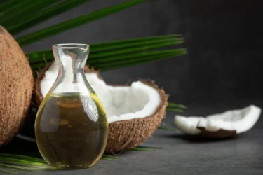
Coconut is a simple fruit with multiple gifts to offer! It is also known as coco, nariyal, coco-da-bahia, etc. The scientific name of the coconut tree is Cocos nucifera (L.), and it belongs to the family Arecaceae. The plant originated from Southeast Asia and islands between the Pacific and Indian Ocean; it was brought to India and East Africa. The coconut palm is either utilized as a whole or in sections to produce milk, husk or the widely used coconut oil. Coconut oil is prepared by pressing dried coconut meat (copra) or fresh coconut meat. The oil prepared using fresh coconut meat is known as virgin coconut oil, and the one prepared using dried coconut meat (copra i.e. the dried brown covering) is called refined coconut oil. Coconut oil has benefits much more than you can expect. Here are a few benefits of virgin coconut oil and some special considerations to be kept in mind if you plan to include coconut oil in your diet1,2.
Coconut oil contains various nutritional components including bioactive compounds such as that are given below (Value per 100 g)3:
You can mix several oils with coconut oil to increase its efficacy when used for hair-related issues such as baldness, hair loss, dandruff, etc. Oils such as castor, amla, neem, and almond are great sources of various nutrients like vitamins, antioxidants, protein, etc. These properties add to the goodness of coconut oil12.
Dr. Siddharth Gupta, B.A.M.S, M.D (Ayu)
The consumption of coconut oil shows numerous scientifically proven properties. Some of these properties are mentioned below:
Some of the potential benefits of coconut oil are described as follows:

Coconut oil prevents damage to various hair types. Rele et al. conducted a review4 in 2003 to assess the effect of different treatments on hair. This study concluded that, among all other treatments, coconut oil was the only oil found to reduce protein loss for damaged and undamaged hair types. In addition, coconut oil is a triglyceride of lauric acid, has a high affinity for hair proteins, that can penetrate hair shafts due to its low molecular weight. This indicates that coconut oil may have a positive impact on hair. However, we need more studies to support these claims4.

Coconut oil is a medium-chain fatty acid known to have several benefits. Teng et al. conducted a systematic review5 in 2020 to assess the effects of coconut oil consumption on lipid profile. The summary estimate of 12 meta-analysis studies showed that consumption of coconut oil increased high-density lipoprotein (HDL- good cholesterol) and low-density lipoprotein or bad cholesterol. A better lipid profile is demonstrated with the use of virgin coconut oil. This indicates that using coconut oil may improve HDL but can also have a harmful effect by increasing low-density lipoprotein. Therefore, more studies are needed to support the use of coconut oil in humans, especially those focused on virgin coconut oil5.

Few studies support the anticancer role of coconut oil. Verma et al. conducted a study6 in 2019 to assess the in-vitro anticancer activity of virgin coconut oil in cancer cell lines in the liver. This study showed positive results for cancer cell lines treated with virgin coconut oil. In addition, the fatty acid component of coconut oil is known to target the liver by portal circulation directly. This indicates that using coconut oil may help manage liver cancers. However, more studies are needed to support these claims6.

Alzheimer’s disease is a prevalent neurodegenerative disease characterised by a decline in cognition. Jose et al. conducted a study7 in 2017 to assess the effect of coconut oil consumption in Alzheimer’s disease. The results of this study showed that the consumption of coconut oil had a positive impact on cognitive function. This suggests that coconut oil may help manage Alzheimer’s disease by improving cognition. However, more studies are needed to support these claims7.

The antiviral and antibacterial properties of coconut oil are attributed to the presence of lauric acid. Khairiyah et al. conducted a literature review8 in 2017 on the nutraceutical properties of coconut oil, which suggested that consumption of coconut oil can have a positive impact on gram positive bacterial infections. Imelda et al. conducted a study in 2021 to assess the effect of virgin coconut oil on Covid-19 patients. The results of this study support the use of virgin coconut oil by reducing C-reactive protein levels that may help manage viral infections like Covid-19. Additionally, the presence of caprylic acid and lauric acid may help in managing fungal infections. The above-mentioned studies indicate that consumption of coconut oil may help in managing bacterial, viral and fungal infections. However, more studies are needed to support these claims8,9.
Some studies show that coconut oil contains lauric acid that possesses antimicrobial activities. Because of these properties, using coconut oil to aid minor wounds might be beneficial13.
Dr. Rajeev Singh, BAMS

Though there are studies that show the benefits of coconut oil in various conditions, these are insufficient and there is a need for further studies to establish the true extent of the benefits of coconut oil on human health.
It can be used in the following ways:
You must consult a qualified doctor before taking coconut oil. Do not discontinue or replace an ongoing treatment of modern medicine with coconut oil without consulting a qualified doctor.
A few side effects related to the consumption of coconut oil include the following9:
However, if you experience any adverse reactions to coconut oil, it is advised to discontinue its intake and immediately contact a doctor or your Ayurvedic physician who has prescribed it to you. They will be able to guide you appropriately for your symptoms.
Eating coconut oil is okay if taken in moderate amounts. However, general precautions must be followed in the following conditions:
Also Read: Walnut Oil: Uses, Benefits, Side Effects By Dr. Smita Barode
There are no significant interactions of coconut oil with other drugs. However, you must always seek the advice of your Ayurvedic physician about the possible interaction of coconut oil with other drugs, and follow the prescription thoroughly, as they will know your health condition and other medications you are taking.
The scientific name of coconut is Cocos nucifera (L.), and it belongs to the family Arecacea1.
The oil prepared using fresh coconut meat is known as virgin coconut oil, and the one prepared using dried coconut meat (copra i.e., the dried brown covering) is called refined coconut oil2.
Studies show a positive result between the consumption of coconut oils and gingivitis, attributed to the presence of polyphenols which show an anti-inflammatory effect. However, more studies must be done to support this claim in humans. Therefore, it is advised to consult a doctor for proper treatment of gingivitis.
Yes, in-vitro studies show that consumption of coconut oil may have a positive impact on liver cancers; however, more studies are needed to ensure this. Therefore, it is advised to consult a doctor for a proper treatment of liver cancer and do not consider consumption of coconut oil as an alternative to modern medicine5.
Consuming coconut oil in excess can result in headaches, nausea, swollen glands, dizziness, chills and loose stools9.
Disclaimer: The information provided here is for educational/awareness purposes only and is not intended to be a substitute for medical treatment by a healthcare professional and should not be relied upon to diagnose or treat any medical condition. The reader should consult a registered medical practitioner to determine the appropriateness of the information and before consuming any medication. PharmEasy does not provide any guarantee or warranty (express or implied) regarding the accuracy, adequacy, completeness, legality, reliability or usefulness of the information; and disclaims any liability arising thereof.
Links and product recommendations in the information provided here are advertisements of third-party products available on the website. PharmEasy does not make any representation on the accuracy or suitability of such products/services. Advertisements do not influence the editorial decisions or content. The information in this blog is subject to change without notice. The authors and administrators reserve the right to modify, add, or remove content without notification. It is your responsibility to review this disclaimer regularly for any changes.
 8
8
 0
0
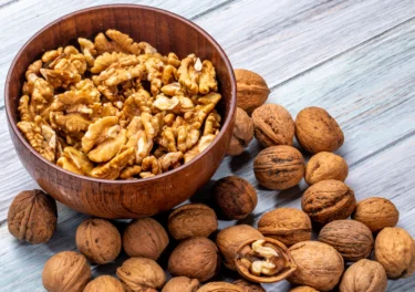
Walnuts are essential for the body to obtain the vitamins and minerals it wants. It is scientifically known as Juglans regia L. and is a member of the Juglandaceae family. Walnut oil is prepared from dried and pressed walnut seeds by cold pressing extraction method. It is also termed ‘akhrot ka tel’ in Hindi. The Romans called it ‘Jovis Glans,’ which means ‘Jupiter’s King Fruit’. There are 18 species of walnut worldwide, but because of its size and sweetness, the Anatolian walnut (Iranian walnut or English walnut) is the most grown commercially. It is grown in India, China, Japan, the United States, Canada, and Central America1,2. Let’s read and find out more about walnut oil benefits and properties.
Did you know?
100ml of walnut oil possesses the following nutrients3
Vitamins present in the walnut oil2:
Walnut oil is a rich source of essential fatty acids, especially alpha-linolenic acid, oleic acid and linoleic acid. Walnut oil also contains proteins, polyphenols, ellagic, malic and gallic acid and phytonutrients like zinc, calcium, iron, potassium, magnesium, phosphorus, copper and selenium2.
Following are the walnut oil properties2:
Omega-3 fatty acids, which are frequently lacking in the diet, are abundant in walnut oil. However, I recommend its use sparingly, much like other cooking oils, to prevent putting too many extra calories in your diet and posing health hazards. For instance, walnuts have the potential to produce a serious allergic response in persons who are allergic to nuts. If they consume walnut oil, some people who are sensitive to peanuts might develop an allergic response7.
Dr. Rajeev Singh, BAMS
Following are the walnut oil uses:

Walnut oil contains monosaturated and polyunsaturated fatty acids and alpha-linolenic acid. It may balance insulin and act against diabetes. In a study4 by Nezhad et al., 2016 eating walnut oil for three months lowers fasting blood sugar and haemoglobin A1c (HbA1c) levels and maintains blood glucose levels. HbA1c is a blood sugar level measurement for the previous two or three months. This study also showed that lowered cardiovascular problems of type-2 diabetes without changing the weight or blood pressure. Thus, walnut oil may show an anti-diabetic effect4. However, diabetes is a serious condition; it can cause severe side effects. So, please consult the doctor for better health results.

Walnut oil contains plant sterols called phytosterols, omega-3 fatty acids and unsaturated fatty acids. These compounds in walnut oil may lower cholesterol absorption into the body. It may also lower blood triglyceride levels. As a result, it may lower total cholesterol levels and low-density lipoprotein (LDL) cholesterol. This way, it may help lower cholesterol in the body2. Even so, more research is required to determine the effect of walnut oil on cholesterol. Kindly consult the doctor and do not self-medicate.

Multiple sclerosis (MS) is an inflammatory disease related to the central nervous system (CNS). T cells are present in the body and kill the infection-causing cells in the body by releasing cytokines. These cells can cause inflammation and damage to the myelin. The covering that surrounds and protects the nerve fibres is myelin. In MS patients, T helper cells, such as Th1 and Th17 and their cytokines can initiate an attack on the CNS. Walnut oil contains anti-oxidant and anti-inflammatory properties, which may lower Th1 activity in MS patients. Walnut oil may help avoid damage to myelin and nerve fibres in the CNS. As a result, walnut oil may be effective in multiple sclerosis5. Because MS is a severe disorder, please consult a doctor for diagnosis and treatment. Do not attempt to self-medicate.

Walnut oil contains essential fatty acids such as linoleic and linolenic acids, which are necessary for normal skin function. It may have moisturising properties and also anti-oxidant properties. In addition, it may assist in regulating trans-epidermal water loss, which is the amount of water that evaporates from the skin. In this way, walnut oil might be beneficial for skin problems. Just like it may benefit dry skin, it may be useful for dry hair and dandruff-prone scalp. As a result, walnut oil may be used for sunburn, skin ulcers and itchy scalp2. However, you must consult a doctor before using walnut oil because it may cause side effects.
According to Liao6 et al., 2020 walnut oil regulates the activity of cholinergic receptors, which is related to regulating physiological activities like attention, memory, learning, and stress response in mice. It may reduce oxidative stress, which is generated by an imbalance between the creation and accumulation of reactive oxygen species (ROS) in cells. It is the ability to detoxify these reactive products. Another study found that eating walnut oil may lower the chance of age-related disorders. As a result, walnut oil might help reduce memory loss in Alzheimer’s patients6. However, walnut oil and its effects on Alzheimer’s disease require more research. Kindly consult the doctor, as Alzheimer’s disease is a serious condition. Do not try to self-medicate.

Phytonutrients and monounsaturated fats in walnut oil may reduce inflammation and help protect the heart. It may also benefit the cardiovascular system by lowering cholesterol and reducing inflammation. More research is in need to determine the effects of walnut oil on cardiovascular diseases2. As walnut oil may cause side effects, kindly consult a doctor for diagnosis and treatment. Do not self-medicate.

The omega-3 fatty acids (alpha-linolenic acid) in walnut oil may reduce inflammation and lower the risk of developing breast, colon and prostate cancer. More research is required to prove the effects of walnut oil on cancer2. As cancer is a severe condition, it may cause severe side effects. Please consult a doctor and do not self-medicate.
Though there are studies that show the benefits of walnut oil in various conditions, but these are insufficient and there is a need for further studies to establish the true extent of the benefits of walnut oil on human health.
There are many calories in walnut oil. To avoid unintended weight gain, I suggest you use walnut oil carefully whether you use it in your cooking, to pour it over salads, or blend it into a sauce. Obesity may lead to major medical concerns including cardiovascular disease, some kinds of cancer, and other health issues7.
Dr. Siddharth Gupta, BAMS, MD (Ayu)
Because of its numerous advantages, usage of vegetable oils is being replaced by walnut oil2. So, let’s look at how to use walnut oil:
You must consult a qualified doctor before taking any herbal supplements. Do not discontinue or replace an ongoing treatment of modern medicine with an ayurvedic/herbal preparation without consulting a qualified doctor.
Walnut oil might cause allergies in some individuals. Therefore, you must ensure that they are not allergic to walnut oil before eating and applying it5. However, if any negative side effects occur while using it, seek immediate treatment. Consult the ayurvedic physician who prescribed it to you; they will be able to identify and solve the problem.
There haven’t been any reports on walnut oil safety precautions. Also, there are no specific precautions to take before its application and consumption in pregnant women, children and the elderly. However, consult a physician before its usage in any form.
Also Read: Matki (moth beans): Uses, Benefits, Side Effects, and More By Dr. Smita Barode
There have been no known adverse drug interactions with walnut oil. However, additional research is necessary for this topic to ensure that it does not interfere with other medications and is safe for use. As a result, your ayurvedic physician’s advice must be carefully followed, as their prescription is based on your health condition.
Also Read: Canola Oil: Uses, Benefits, Side Effects by Dr. Smita Barode
Walnut oil contains phytonutrients such as selenium, phosphorus, magnesium, zinc, iron, and calcium. Those may help in the management of the diseases such as diabetes, cancer, heart disease, and hypertension. They may also act as precursors and catalysts for hormones in the body. In addition, it may improve metabolism, the nervous system, digestive and brain function, and energy production2. Even so, please consult the doctor as walnut oil may cause adverse effects.
No, but additional research is needed to determine the effect of walnut oil on migraine. Please consult a doctor for better health results.
No. However, more research is required to determine the benefits of walnut oil for the thyroid. Kindly consult a doctor for proper diagnosis and treatment.
Walnut oil may be topically applied for the treatment of dandruff and dry hair1. Although, more research on the effect of walnut oil on hair is required. Kindly consult a doctor.
Walnut oil may interact with drugs and change their activity. However, no such information is available. More research is needed on this topic. Please check with your doctor before consuming walnut oil, primarily if you are on any medication.
Disclaimer: The information provided here is for educational/awareness purposes only and is not intended to be a substitute for medical treatment by a healthcare professional and should not be relied upon to diagnose or treat any medical condition. The reader should consult a registered medical practitioner to determine the appropriateness of the information and before consuming any medication. PharmEasy does not provide any guarantee or warranty (express or implied) regarding the accuracy, adequacy, completeness, legality, reliability or usefulness of the information; and disclaims any liability arising thereof.
Links and product recommendations in the information provided here are advertisements of third-party products available on the website. PharmEasy does not make any representation on the accuracy or suitability of such products/services. Advertisements do not influence the editorial decisions or content. The information in this blog is subject to change without notice. The authors and administrators reserve the right to modify, add, or remove content without notification. It is your responsibility to review this disclaimer regularly for any changes.
5
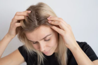
Do you get a feeling of constant itch on your scalp? Does it make you uncomfortable? If yes, you might have a problem that needs your immediate attention. An itchy scalp is a condition where you have a strong feeling of irritation in your scalp that makes you want to scratch badly. The medical term for itchy scalp is Scalp pruritus. It is a distressing, although common symptom that is considered a challenging diagnosis. It is commonly associated with psoriasis and seborrheic dermatitis. The itchy scalp condition is not well-studied1,2.
It is very unpleasant to scratch your head in public. Therefore, we have a few natural remedies for itchy scalp, which you can try in the comforts of your home. So, continue reading to find natural solutions for itchy scalp.
Did you know?
Itchy scalp may be caused due to several reasons. Most common causes include:
You might experience an itchy scalp without dandruff and other situations where your head has lice or probably dermatitis, which needs attention from a dermatologist. This situation might need an intervention with a topical agent like steroids, with treatment sometimes for a period of around four to six months.
Dr Ashish Bajaj, M.B.B.S, M.D. in Clinical Pharmacology and Toxicology
Although itchy scalp is a diagnosis, it is also a symptom that helps diagnose other scalp diseases. Most common symptoms that are associated with itchy scalp are given below:
There are various natural home remedies for itchy scalp that you may try.
Walnut leaves have been used in traditional medicine. It may be a suitable remedy for itchy scalp. The leaves may be useful for external applications such as itchy scalp, dandruff, as an emollient (soothing agent), and to soothe itching in skin disorders3. A decoction of walnut leaves might be a beneficial remedy for itchy scalp. It is made by boiling dry walnut leaves on low heat for 15-20 minutes. Then strain the leaves and use the liquid for rinsing the scalp or add it to your bathwater.

Amla, also called Indian gooseberry, is a popular Ayurvedic herb. Amla contains a vast array of antioxidants, vitamins and minerals. It may also have beneficial properties such as antimicrobial, anti-inflammatory activities and cooling effects. Amla oil, amla juice and dried amla may be used as a possible home remedy for itchy scalp. Amla oil may help reduce scalp irritation and other infections related to the scalp3. Amla powder may provide instant relief to your itchy scalp. Amla powder is made by grinding dried amla fruit. Make a thin paste by mixing this powder with some water. Then apply it to your scalp for 15-20 minutes and wash it off.
Shikakai has been used for managing dandruff, strengthening hair follicles, etc. It has a low pH value and may be useful for scalp and hair conditioning as well as cleansing. Shikakai powder may be a beneficial home remedy for itchy scalp. A paste made from shikakai powder using water can be applied to the scalp. It may act as a great conditioner and reduce the itch and dandruff3.
Coconut oil is a great source of antioxidants, vitamin E and K, minerals and lauric acid. It is believed that coconut may have cooling properties because of which it is used by people with Pitta Dosha. It may also have beneficial properties such as antioxidant, antiviral, antibacterial and antifungal properties. Coconut oil may be a good remedy for itchy scalp. It might be useful to fight dryness, reduce dandruff and eliminate scalp infections and fungus-forming units3. You may directly apply coconut oil to the affected regions of the scalp. Let it sit for some time and then rinse it off.
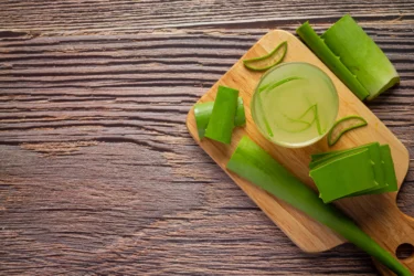
Aloe vera may also be considered one of the most useful home remedies for itchy scalp. Aloe is collected as a dried juice by cutting the base of the leaves. It may be a beneficial conditioner for hair and scalp. It may benefit in reducing the itching of the scalp and dandruff and may help condition the hair. The prominent benefiting component present in Aloe vera is called aloenin3. Aloe vera gel may be beneficial for itchy scalp. To make aloe vera gel take a leaf and remove the covering of the leaf. Next, scoop the gel with the help of a spoon, put it in a blender and blend it for a few seconds to make a frothy, liquefied gel. Finally, you can apply this gel to your scalp to relieve the itchy scalp.
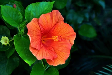
Hibiscus may be used as a home remedy for itchy scalp. It contains vitamins A and C and iron. It may have beneficial properties like antioxidant, antibacterial and anti-inflammatory properties. Hibiscus herbal preparations may be beneficial for itchy scalp. A herbal mask can be prepared using hibiscus flowers and leaves. It may condition your scalp and hair as well as reduce dandruff3.
Take 3 to 4 hibiscus leaves and one flower and grind them together to make a fine paste. You can mix it with any of these (yoghurt, aloe vera gel, almond oil, olive oil or coconut milk). Apply the paste to the scalp for an hour or more and rinse it off with water.
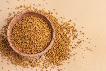
Fenugreek or methi may be a natural, secret ingredient for hair and scalp health. It contains potassium, iron, vitamin C, lecithin and proteins. It might reduce dandruff and may provide a soothing effect to the dry and itchy scalp3. Fenugreek paste may help manage the itchy scalp. To make methi paste, soak 1-2 tablespoons of methi seeds in water overnight. Next morning, grind these seeds into a fine paste using the same water. You can apply this paste to your dry, itchy scalp to reduce the itch.
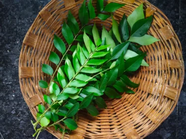
Curry leaves contain a high amount of minerals like iron and antioxidants such as vitamins C, A, E and folic acid. It may also have antibacterial properties that might help reduce scalp infection. Curry leaves may have a nourishing effect on both hair and scalp3.
Curry leaves paste may be beneficial for your itchy scalp. To make curry leaves paste, take a handful of curry leaves and mix them with yoghurt to achieve a paste consistency. Apply this paste to your scalp and allow it to rest for one hour. Then rinse thoroughly with cold water. Repeat this procedure 2 to 3 times a week for best results.
Also Read: Home Remedies For Hair Thinning
Castor oil may have antimicrobial, antifungal and anti-inflammatory properties. It contains ricin and resinoleic acid that might nourish the scalp and hair. It may also have the ability to enhance blood circulation. Castor oil might help combat dryness and dandruff, which would be beneficial for itchy scalp3.
Though studies show the benefits of the given herb and home remedies for itchy scalp conditions, these are insufficient. Therefore, there is a need for large-scale human studies to establish the true extent of the benefits of these home remedies on itchy scalp. Thus, these should only be used cautiously and never as a substitute for medical treatment.
Lemon juice is one of the most popular scalp itching home remedies. It has antibacterial properties that prevent the yeast Malassezia from causing dandruff. Also, its acidic properties help soften the already existing flakes and calm down the inflammation on the scalp.
Dr. M.G. Kartheeka, MBBS, MD(Pediatrics)
Also Read: How To Increase Melanin In Hair Naturally
An itchy scalp might be indicative of serious conditions, so it is important to treat it properly. Therefore, people should consult a doctor if the conditions mentioned below are seen.
You must not rely on home remedies alone for the treatment of itchy scalp. You should always consult a dermatologist or any other qualified doctor if the symptoms do not improve with home remedies.
Also Read: Effective Home Remedies For Dry Scalp
Itchy scalp is medically termed Scalp pruritus. It is recognised by constant irritation and a distressing feeling on the scalp. Itchy scalp is a challenge to diagnose medically. Various factors such as the presence of lice in the head, fungal infection, dandruff, inflammation of the scalp and skin diseases like psoriasis and seborrheic dermatitis cause itchy scalp. However, there are several home remedies for itchy scalp that you can use. Various herbs, including amla, walnut, shikakai, coconut oil, castor oil, neem, tulsi, aloe vera, hibiscus, fenugreek, curry leaves, etc. might be beneficial home remedies for itchy scalp. However, if the condition persists for longer, seek medical attention to avoid further complications.
Also Read: Effective Home Remedies for Silky Hair
The causes of the itchy scalp, such as head lice, are difficult to manage because of the easy spread of head lice. The spread is prevented by avoiding sharing hairbrushes and combs. For chronic conditions such as psoriasis, dandruff or seborrheic dermatitis, a regular doctor’s appointment and treatments according to the conditions are required. A medicated shampoo prescribed by a pharmacist might help with this. Maintaining personal hygiene is essential for treating tinea infection1.
According to doctor’s recommendations, medicated anti-dandruff shampoos, antifungal medicine for tinea capitis, moisturising creams and ointments, head lice treatment, corticosteroid creams for seborrheic dermatitis and psoriasis medications are few of the treatment options for itchy scalp1. However, people should use these treatments only after consultation with a doctor and if prescribed.
Thyme might be beneficial for reducing dandruff and may be used in scalp rub3. However, prior to using thyme as a home remedy for itchy scalp, people should consult a doctor.
Tulsi contains the antioxidant vitamin K. It may be an essential herb for eliminating bacterial and fungal infections, which may help with treating an itchy scalp3. However, using Tulsi to treat an itchy scalp is not recommended without consulting a doctor.
Yes. Flaxseed is a rich source of antioxidants and fatty acids. It may help remove the dead cells from the scalp. Applying flaxseed paste to your hair and scalp may act as a good moisturiser3. However, people should not use flaxseeds to self-medicate without consulting a physician.
Neem oil or neem extract may be used for treating psoriasis and dandruff. However, it is important to refrain from using neem without consulting a doctor.
1. Health direct [Internet]. Itchy scalp. 2020 [cited 2022 Jul 11]. Available from: https://www.healthdirect.gov.au/itchy-scalp
2. Bin Saif G, Ericson M, Yosipovitch G. The Itchy scalp – scratching for an explanation. Experimen dermato. 2011;20(12):968. Available from: https://www.ncbi.nlm.nih.gov/pmc/articles/PMC3233984/pdf/nihms-331222.pdf
3. Kolekar Y, Tamboli, Firoj, More H, Mulani S, Mali N. Medicinal plants used in cosmetics for skin and hair care. Intern J of Pharmaceu Chem and Analy. 2021;8(2):36–40. Available from: https://www.ijpca.org/html-article/14382
4. Cleveland Clinic [Internet]. 5 Causes (and Fixes) for Itchy Scalp. 2020 [cited 2022 Jul 20]. Available from: https://health.clevelandclinic.org/itchy-scalp-5-common-problems-and-fixes/
Disclaimer: The information provided here is for educational/awareness purposes only and is not intended to be a substitute for medical treatment by a healthcare professional and should not be relied upon to diagnose or treat any medical condition. The reader should consult a registered medical practitioner to determine the appropriateness of the information and before consuming any medication. PharmEasy does not provide any guarantee or warranty (express or implied) regarding the accuracy, adequacy, completeness, legality, reliability or usefulness of the information; and disclaims any liability arising thereof.
Links and product recommendations in the information provided here are advertisements of third-party products available on the website. PharmEasy does not make any representation on the accuracy or suitability of such products/services. Advertisements do not influence the editorial decisions or content. The information in this blog is subject to change without notice. The authors and administrators reserve the right to modify, add, or remove content without notification. It is your responsibility to review this disclaimer regularly for any changes.
17
3
Have you seen a snake like appearing vegetable? If not, then you might be astonished to know that there is a greenish white (when immature) long and cylindrical vegetable that turns dark red (when mature)1. It is known as snake gourd, long tomato, tomato gourd or viper gourd in various countries. It is commonly grown in Asian countries like India, Sri Lanka, Peninsula, Philippines and Malaysia. The plant is a creeper and belongs to the family Cucurbitaceae2.
Snake gourd is scientifically known as Trichosanthes cucumerina. In India, it is referred to by a different name in region.
For example, it is known as Pudalankaai in Tamil
Snake gourd has been used in Ayurveda and Siddha since ancient times. The entire plant of snake gourd might be beneficial as the roots, leaves, fruits and seeds all have potential benefits against various ailments. In traditional medicine systems of Ayurveda and Siddha, it is used in polyherbal preparations1.
Snake gourd has a high nutritional value with the following nutrients present in it.
Snake gourd might have the following properties:
Snake gourd belongs to the pumpkin family (Cucurbitaceae). Due to its long, slender and curving-like snake, it is known as a ’snake gourd’. It can easily reach up to 1.5 m in length3.
Dr. Siddharth Gupta, B.A.M.S, M.D (Ayu)
There are very few studies to know the potential benefits of snake gourd. The existing studies show that snake gourd might have the following potential uses for human health.

A study2 was conducted on Albino rats to explore the antidiabetic potential of snake gourd. In this study2 it was found that the ethanolic extract of snake gourd might have a positive effect on the blood sugar level. Snake gourd might help in increasing the glucose tolerance of the body through absorption into surrounding tissues. Therefore, it might be helpful for diabetes. However, more research on humans is required to prove these effects. Moreover, it is advised that you consult a doctor for proper diagnosis and treatment for serious conditions such as diabetes2.

In the study2 conducted by Arawwawala proved that snake gourd might help protect the stomach from damage from substances such as ethanol and indomethacin. The snake gourd extract might help increase the protective mucous layer of the stomach, may interfere with the activities causing damage to stomach cells (antihistamine activity) or may help decrease the acidity of the gastric juice. However, further research is required to prove these claims.

As previously mentioned, the whole plant of snake gourd was used in traditional medicine. The protective activity towards liver might be attributed to the whole plant. The ethanol extract of the whole plant might have potential to protect the liver from carbon tetrachloride induced hepatotoxicity2. However, this is not a substitute for a consultation with a health professional2.

It was seen in animal models that snake gourd might have the potential to help relieve inflammation. In an experiment done by Kolte RM et al. in 1997, the hot root extract of snake gourd showed the potential to alleviate inflammation seen in animal models. However, further research on humans is required to prove these effects2.
The root extract and the fruit extract of snake gourd were tested for their effect against cancer cells. This study conducted by Kongtun S et al. in 1999 showed that both these extracts of snake gourd showed the potential to be effective against the growth of breasts, lungs and colon cancer. Further, it was seen that the root extract showed greater potential than the fruit extract. Therefore, snake gourd might have the potential to help against cancer. Please note that cancer is a serious condition and must be properly diagnosed and treated by a doctor. Kindly visit a doctor and do not self-medicate2.

A study2 showed that snake gourd might be effective against larva of certain insects. Therefore, it might help with certain infections caused by insects.
Traditionally, snake gourd has been used to cure certain ailments. So, it might have the potential to help with fever, headache, alopecia, skin allergy, colic (sudden or abrupt stomach pain), boils, diarrhoea, haematuria (blood in urine), bronchitis, malaria, etc2.
Though there are studies that show the potential uses of snake gourd in various conditions, but these are insufficient and there is a need of further studies to establish the true extent of benefit of snake gourd on human health.
Snake gourd is a vegetable that is usually cooked before eating. It can be had in form of curry, stir fried or added to soups.
You must consult a qualified doctor before taking any herbal supplements. Do not discontinue or replace an ongoing treatment of modern medicine with an ayurvedic/herbal preparation without consulting a qualified doctor.
Due to its rich vitamin and mineral contents, snake gourd might stimulate new hair growth. It might also help in strengthening weak hair follicles. Additionally, snake gourd might also help in reducing dandruff4.
Dr. Rajeev Singh, BAMS
Although snake gourd is relatively well consumed as a vegetable, the side effects of snake gourd are yet to be studied. If you notice any unusual symptoms after eating it, you should consult a doctor immediately.
Also Read: Kale: Uses, Benefits, Side Effects By Dr. Smita Barode
It is advised that you follow the general precautions such as looking for the quality of snake gourd before its consumption. It is important to take extra caution along with doctor’s advice before giving snake gourd to pregnant women, breastfeeding mothers, elderly individuals and children.
Also Read: Turmeric (Haldi): Uses, Benefits, Side Effects, and More!
Snake gourd might react with certain drugs and have adverse reactions. However, there is insufficient data regarding the drug interactions. Therefore, it is best that you consult a doctor, especially if you are undergoing any treatment.
Also Read: Pumpkin (Kaddu): Uses, Benefits, Side Effects By Dr. Rajeev Singh
Snake gourd may be helpful in diabetes, for stomach against inflammation and parasitic infestations, etc. However, the potential uses of snake gourd need to be further investigated. Therefore, please consult a doctor for the mentioned conditions and do not self-medicate1,2.
Nutrients present in snake gourd are carbohydrates, proteins, fats and water. The predominant minerals present in snake gourd are potassium and phosphorous. It contains vitamins A and E. It also contains various bioactive components like carotenoids, flavonoids, etc1.
As seen in several studies, snake gourd might increase the glucose tolerance, may increase the uptake of glucose by surrounding cells, might have insulin-like actions. Therefore, it may be helpful in diabetes. However, please consult a doctor for proper diagnosis and treatment of diabetes. Do not self-medicate.
There are no reports regarding the benefits of snake gourd for skin. Therefore, there is need for more research.
Snake gourd is known by different names in different languages. It is known as Chachinda in Hindi. Some of its other names are, Pudalankaai in Tamil, Potlakaaya in Telugu, Chichinga/Chichingae in Bengali, Padabali in Gujarati, Galatori in Punjabi, Aduvulakaayi in Kannada and Padavalanga in Malayalam.
Disclaimer: The information provided here is for educational/awareness purposes only and is not intended to be a substitute for medical treatment by a healthcare professional and should not be relied upon to diagnose or treat any medical condition. The reader should consult a registered medical practitioner to determine the appropriateness of the information and before consuming any medication. PharmEasy does not provide any guarantee or warranty (express or implied) regarding the accuracy, adequacy, completeness, legality, reliability or usefulness of the information; and disclaims any liability arising thereof.
Links and product recommendations in the information provided here are advertisements of third-party products available on the website. PharmEasy does not make any representation on the accuracy or suitability of such products/services. Advertisements do not influence the editorial decisions or content. The information in this blog is subject to change without notice. The authors and administrators reserve the right to modify, add, or remove content without notification. It is your responsibility to review this disclaimer regularly for any changes.
11
4
If you are searching for a healthy drink to kick start your day, look no further! Packed with numerous health benefits, neem juice can be your choice for a healthier and better future.
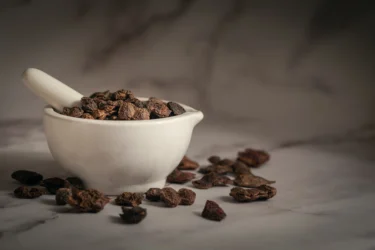
As people become more mindful of their health concerns, more and more people have started relying on herbal juices. Neem juice is a bitter juice obtained from crushing the leaves of the neem tree. Neem, scientifically known as Azadirachta indica, belongs to the Meliaceae family. The neem plant is commonly found in India, Bangladesh, Nepal and Pakistan. Neem has been widely used in traditional systems of medicine such as Chinese medicine, Unani medicine and the Ayurvedic system of medicine1.
Effective properties of many plants are being researched for disease management because of their fewer side effects. Neem juice contains numerous phytochemicals that possess functional properties against diseases. The effective properties of neem leaves are very well documented1.
If you want to learn about bitter neem juice health effects, continue reading!
Some nutrients found in neem leaf are minerals like calcium and phosphorous2. Neem juice also contains plenty of phytochemicals such as
Owing to the hist of phytochemicals it contains, neem juice may show the following properties for human health.
Neem juice possesses many valuable properties because of which it has potential uses for many health conditions. However, most of these uses have been observed in lab-scale trials using neem leaf extract. Neem juice may show properties similar to leaf extract and may show effects against several disease conditions. However, more research is required to consolidate its uses in people. Therefore, you are advised to consult a physician before using neem juice on your own as a remedy for any serious medical condition.

Neem leaf extract could help promote wound healing as observed during several animal studies. This wound healing property was observed on rats. Using neem juice may show properties similar to extract. Therefore, you may use neem juice to help accelerate wound healing1. However, if you are suffering from any wound, you still need to take complete wound care to prevent the wound from getting infected. Avoid using neem juice without consulting with a doctor first.
Neem extract has been evaluated for its antidiabetic activity in lab studies. Neem extract may show positive effects on blood glucose and help reduce the activity of the glucosidase enzyme (responsible for breaking complex carbohydrates into glucose). Neem leaf extract also showed activity against diabetes mellitus1. These activities may help manage diabetes. However, if you have diabetes, avoid using neem juice or any other herbal remedy without consulting your healthcare provider first.
Neem leaf extract showed good potential as a liver protectant. The positive effects of neem leaf extract against liver toxicity and damage were observed during animal studies. Neem leaf extract could show positive effects during the trial by restoring the levels of liver enzymes1. These effects may point in a positive direction. However, you must reach out to your healthcare provider in case of any liver problems. Trying to treat liver problems with herbs and tonics without consulting a doctor may worsen the situation.
As neem has hepatoprotective, gastroprotective, and antispasmodic properties, neem juice might be beneficial for maintaining digestive health. Drinking neem juice might promote bowel movement, improve liver function and might relieve constipation6.
Dr. Rajeev Singh, BAMS

Neem leaf extract has shown antibacterial activity against several foodborne bacteria. The activity against bacterial growth has also been observed in other lab trials. Neem leaf extract has also shown activity against the growth of several fungal species during lab studies. In addition to activities against bacteria and fungi, the potential use of neem extract against the growth of coxsackievirus has been observed1. Neem juice may have these properties as well. All these activities were observed during lab studies. If you are suffering from any infection, bacterial, viral or fungal, you must get treated by a healthcare provider and receive proper treatment as larger studies are needed to confirm efficacy in humans.
Neem leaves are proven to have antifungal properties. Because of this, the juice extracted from its leaves might be beneficial for reducing dandruff from the scalp and maintaining healthy hair and a clean scalp5.
Dr. Siddharth Gupta, B.A.M.S, M.D (Ayu)
Neem leaves possess potent antioxidant and anti-inflammatory activities. Free radicals in the body often lead to chronic diseases in the long run. Neutralizing these free radicals may help avoid the onset of many health conditions. The antioxidant properties of neem leaf may help destroy or neutralize the free radicals generated in the body. In addition, the anti-inflammatory properties of the neem leaf may also help with inflammation and swelling in the body1.
Though some studies show the benefits of neem juice in various conditions, these are insufficient, and more research to establish the true extent of the benefits of neem juice on human health.
To make fresh neem juice, you can clean some fresh leaves with water. Grind the leave and add water to it to make neem juice. You may consume raw neem juice to partake its health effects3.
You must consult a qualified doctor before taking neem juice or any herbal supplements. Likewise, do not discontinue or replace an ongoing treatment of modern medicine with an ayurvedic/herbal preparation without consulting a qualified doctor.
Various studies are reporting the liver toxicity of neem. Therefore, people with liver problems need to be careful while taking neem juice or herbal juice4.
Using neem juice without consulting with a doctor or physician is not advised. Before taking neem juice, you need to consult a doctor about its potential limitations, side effects and safe dose.
Also Read: Tomato (Tamatar) Juice: Uses, Benefits, Side Effects By Dr. Smita Barode
Here are some precautions you need to take while using neem juice.

There is a lack of sufficient information to support the use of neem juice during pregnancy or breastfeeding. Therefore, it is better to be on the safer side and avoid taking neem without consulting first with your doctor or healthcare provider.

There is a lack of information supporting the use of neem juice. Therefore, it is better to be on the safe side and avoid its use unless prescribed by a certified doctor or healthcare provider.

People suffering from liver ailments should avoid neem juice, which can lead to liver injury and damage4.
Before taking neem juice or any herbal juice, you need to first consult with a doctor about its limitations and precautions. Avoid using neem juice unless consulted by a doctor or physician.
Also Read: Ginger Juice: Uses, Benefits, Side Effects By Dr. Siddharth Gupta
Because of the various properties of neem leaf, it may show herb-drug interaction.
People who are taking acetaminophen regularly should avoid drinking neem leaf juice. Regular use of acetaminophen with neem leaf extract lead to liver toxicity in rats during lab trials4.
Avoid using neem juice for its properties or against any disease without consulting your doctor. Also, if you are taking some medicine, always consult your doctor about any herbs or juices you need to avoid. It’ll help you prevent serious health consequences.
Also Read: Lauki Juice: Uses, Benefits, Side Effects and More!
Neem juice may show properties against diseases like diabetes, liver problems and microbial infections. Neem juice may also show wound healing and anti-inflammatory and antioxidant properties1. However, before using neem juice for these effects, you are advised to consult with your healthcare provider. Avoid using herbal supplements without consulting with your doctor.
Intake of neem juice may lead to liver toxicity. There are several reported cases of neem extract causing liver toxicity4. If you have a weak liver or any liver problems, you are advised not to use neem juice. Use this juice only after consulting with a doctor.
Neem juice may show anti-inflammatory and antioxidant properties. It may help with inflammation and swelling of acne on the skin. It may also help purify the blood1,3. However, you can contact your skin doctor if you are suffering from any skin condition. Avoid using any herbal supplement without a doctor’s consultation.
Several lab studies are reporting the positive effects of neem juice on diabetes1. However, these studies are few and more studies are required to support neem juice use by people with diabetes. You should not use herbal supplements for serious health conditions like diabetes without consulting your doctor.
Disclaimer: The information provided here is for educational/awareness purposes only and is not intended to be a substitute for medical treatment by a healthcare professional and should not be relied upon to diagnose or treat any medical condition. The reader should consult a registered medical practitioner to determine the appropriateness of the information and before consuming any medication. PharmEasy does not provide any guarantee or warranty (express or implied) regarding the accuracy, adequacy, completeness, legality, reliability or usefulness of the information; and disclaims any liability arising thereof.
Links and product recommendations in the information provided here are advertisements of third-party products available on the website. PharmEasy does not make any representation on the accuracy or suitability of such products/services. Advertisements do not influence the editorial decisions or content. The information in this blog is subject to change without notice. The authors and administrators reserve the right to modify, add, or remove content without notification. It is your responsibility to review this disclaimer regularly for any changes.
9
4
The ash gourd plant is an annual trailing vine. It is scientifically known as Benincasa hispida (Thunb.) and belongs to the family Cucurbitaceae. It has a unique melon-like fruit that is often eaten for its medicinal and functional properties. Ash gourd plant grows in warm, humid tropical climates and is cultivated in countries of South East Asia, including India, Japan, China, Myanmar, Malaysia, Indonesia and Taiwan1.
The ash gourd fruit got its name from the colour of its skin, which is ash, and it is also called wax gourd because of the waxy shine on the skin. It is mainly grown during the rainy season, so people also call it the winter melon. In ancient medical systems, the Ayurvedic preparation of ash gourd, known as ‘Kushmanda’ in Sanskrit, was thought to have medicinal properties. The most famous sweet, ‘Agra ka Petha’, is also prepared using the ripened ash gourd fruit soaked in sugar syrup2. There are several other names for ash gourd that you might have heard of; it is named Donggua in Chinese, Beligo in Indonesia, White gourd, White pumpkin, Ash pumpkin, etc.
Let’s read more about the beneficial properties and potential uses of ash gourd juice.
The essential nutrients of ash gourd fruit are proteins, flavonoids, carotenes, vitamins, minerals, volatile oils, etc. Ash gourd fruit is majorly composed of 96% water; the remaining nutrients are as follows3:
Ash gourd fruit is a common vegetable that might have nutritional and medicinal properties. The bioactive nutrients might show potential benefits in various chronic diseases. The properties of ash gourd juice are5:
Ash gourd is considered a functional food, as all parts of the fruit have demonstrated beneficial properties in certain studies5. However, how helpful these are for humans needs to be seen by larger studies. Here are some of its potential uses:
Ash gourd juice benefits people with diabetes, as it is low in calories and carbohydrates with no fat content. It might be a great choice for patients with diabetes due to this nutrient profile. The pulp of the fruit might have anti-diabetic properties contributed by various nutrients. When mixed with honey, the dried powder of the peel of the fruit may help lower blood sugar levels.
A human study6 conducted by Majumdar et al. (2010) observed that ash gourd juice helps reduce blood glucose levels in patients with Type 2 diabetes. However, more studies are required to present the benefits of ash gourd juice for diabetes.

The various properties of ash gourd juice might help in weight loss management. As we know, ash gourd juice is low in calories and fats and thus it may be beneficial to people who want to lose weight. The lipid-lowering properties and high content of dietary fibre of ash gourd juice might help decrease serum cholesterol and lipid levels, which may help lower body fat.
A literature7 review by Waidyarathna et al. (2020) suggests that if you use ground peeled raw ash gourd fruits and seeds with an equal amount of water and some salt, it might be helpful to lose weight. Although this information is insufficient and requires more studies.

The health benefits of ash gourd juice might positively affect people suffering from peptic ulcers (related to the digestive tract and stomach). Ash gourd juice is prepared by shredding the fruit and mixing it with water. Drinking ash gourd juice on an empty stomach may be helpful for peptic ulcers. After consuming the juice, it is best to avoid eating for at least three hours. According to the Ayurvedic medicine system8, ash gourd has been referenced as a valuable medicine for peptic ulcers. This information is age-old and insufficient; therefore, large-scale human studies are necessary to confirm these benefits.

Ash gourd juice may support skin health. The fruit extract used to prepare face cream might be helpful and effective in delaying the deterioration of skin cells with age. A study9 showed that some compounds of the fruit (pulp, peel, seeds) contribute to its antioxidant activity, which may fight ageing-inducing free radicals. It may also decrease oxidative damage and help manage the effects of skin cell degradation. More studies are required to confirm the beneficial effect of ash gourd juice on skin.

Although some studies show the benefits of ash gourd juice in various conditions, these pieces of information are insufficient. Therefore, more studies are required to confirm the benefits of ash gourd juice on human health. Hence, you should always consult a doctor before using ash gourd juice for any medical condition.
Based on my experience, I have come across research4 suggesting that ash gourd may have a potential protective effect on Alzheimer’s disease. Ash gourd showed promise in protecting neurons against oxidative stress, which is believed to contribute to the development of Alzheimer’s disease. This protective effect could be attributed to the presence of vitamin E and β-carotene in ash gourd, which helps counteract oxidative damage.
Dr. Siddharth Gupta, B.A.M.S, M.D (Ayu)
Ash gourd juice contains significant nutrients essential for maintaining good health. It can be used in the following ways:
Before consuming ash gourd fruit or juice, it is better to take the advice of a qualified doctor. They will be the best person to prescribe you the correct way to use it.
Over the years, I have come across research3 suggesting that ash gourd may have an anti-compulsive effect. An anti-compulsive effect refers to the ability of a substance or treatment to reduce or alleviate compulsive behaviours. This effect could be attributed to the presence of tryptophan in ash gourd, which is believed to enhance the production of serotonin, a neurotransmitter that helps regulate mood and behaviour.
Dr. Rajeev Singh, BAMS
While generally safe, ash gourd juice may have some side effects. It is important to be aware of ash gourd juice side effects, as it contains anti-nutritional factors (e.g., phytates, oxalate, etc.), which might reduce the body’s ability to absorb nutrients. A diet containing ash gourd juice may increase the risk of calcium deposition, which might lead to kidney stones11.
Various toxicological studies on animals suggest that ash gourd juice may be safe and might not have any adverse effects. However, if you experience any harmful reaction, immediately contact an Ayurvedic doctor and get appropriate treatment.
In my experience, I have observed that ash gourd extract has shown effectiveness in reducing allergic inflammation. These effects may be attributed to certain compounds present in ash gourd that help to alleviate inflammation10.
Dr. Smita Barode, BAMS, M.S.
Also Read: Excellent Health Benefits Of Sugarcane Juice
Ash gourd is considered safe if it is taken in minimal dosages. However, precautions have to be followed while having ash gourd juice.
We advise you to consult an Ayurvedic physician for a safer dosage form.
Also Read: Raw Papaya: Uses, Benefits, Side Effects and More!
There is not enough information. However, it is recommended that ash gourd juice should not be taken orally along with any mineral drugs.
It is always best to follow the advice of an Ayurvedic physician who will guide and prescribe you a better way to make use of this herbal juice.
Also Read: Carrot Juice: Uses, Benefits, Side Effects and More!
Other vernacular names for ash gourd are Kundur (Malay), Safed Kolu (Gujarati), Neer oosanikai (Tamil), Kumbalam (Malayalam), Boodida gummadikaaya (Telugu), Boodu gumbala (Kannada) and Alupuhul (Sinhalese)3.
Ash gourd juice contains essential minerals necessary for performing vital functions and maintaining water balance in the body; sodium might help to maintain body fluid balance, whereas calcium and potassium might maintain healthy blood pressure. Iron helps to transport oxygen to the brain, zinc nourishes the body, and manganese performs cellular activities.
The dental benefits of ash gourd juice may be involved in managing teeth and gum health. Using ash gourd juice daily for mouth gargling may help maintain your teeth and reduce bleeding gums. However, one should consult a doctor before using ash gourd juice for dental health.
Drinking ash gourd juice might be helpful in weight management due to its low calories. However, this information is insufficient to claim the beneficial effect of ash gourd juice. Hence, more studies are needed.
Yes, ash gourd juice may have antacid properties that might regulate the acidity in the stomach. People might use ash gourd juice for acidity, by boiling it with cow’s milk. However, you should consult an Ayurvedic doctor before using it to control acidity.
Disclaimer: The information provided here is for educational/awareness purposes only and is not intended to be a substitute for medical treatment by a healthcare professional and should not be relied upon to diagnose or treat any medical condition. The reader should consult a registered medical practitioner to determine the appropriateness of the information and before consuming any medication. PharmEasy does not provide any guarantee or warranty (express or implied) regarding the accuracy, adequacy, completeness, legality, reliability or usefulness of the information; and disclaims any liability arising thereof.
67
5
Are you worried about oily and greasy-looking hair? Here are some remedies that can be used at home to keep your hair oil-free and clean. Oily scalp and hair are common problems many people face at some point in their lives. Oily and greasy-looking hair can make people self-conscious. The scalp has more number of sebaceous (oil) glands present, which can contribute to the oiliness of the scalp and hair. Sebaceous glands present on the scalp produce a large amount of sebum (oil), giving you an oily scalp and hair1. Keeping the scalp clean is essential for getting healthy hair. If the presence of excessive oil on the hair and scalp makes you uncomfortable or self-conscious, you can contact your healthcare provider and get the necessary treatment. Do inform them of any past allergies from medications or any treatment that you may have taken. You can also use these remedies to help you eliminate the excess oil on the hair and scalp.
Oil glands are the glands present beneath the skin surface. These glands are more abundant on the scalp and face. Oil glands are formed alongside a hair follicle. Oil glands produce oil, which imparts oiliness to the hair and scalp1.
Several factors govern the secretion of oil on the scalp. These factors are:
Don’t brush your hair too much. While you should brush your hair as needed, over-brushing can cause the hair follicles to produce more oil.
Dr Ashish Bajaj, M.B.B.S, M.D. in Clinical Pharmacology and Toxicology
Signs of oily scalp and hair are the oily and glossy appearance of hair. Other serious symptoms of oily hair and scalp are given below.
Here are some remedies that you can make use of to take care of excessive oiliness of the hair and scalp.
Tulsi leaf paste can be applied to the scalp to clean the hair roots and scalp. It may also soothe the scalp, improve blood circulation, and reduce irritation and dandruff. Irritated and damaged skin produces more oil. It is valuable for keeping the scalp healthy and promoting the growth of hair3. You can use fresh leaves of tulsi to make a paste. Apply this paste to hair and scalp evenly and wash it off with water after some time. Washing the hair and scalp with tulsi paste may help you get rid of oil from the hair and scalp.
Shikakai is often used as a natural cleanser. It has been used traditionally to make herbal shampoos. Shikakai is used as an ingredient in herbal shampoos. It is valuable for hair3. You can use shikakai powder to wash hair and remove the oil from the hair and scalp. Shikakai powder can be used to wash the hair. You can also make shikakai powder to make a hair pack. Mix shikakai powder with some yoghurt to make a paste. Apply this paste as a hair pack. Use this hair pack once a week to get rid of excess oil from hair and scalp.

You can use neem leaves as a cleanser while washing the hair. Neem might help open the clogged pores on the scalp and promote hair growth. Neem is also effective against problems like dandruff, which can be caused by an oily scalp. You can use neem leaf powder to wash your hair3. Take some neem leaf powder and mix water to make a grainy paste. Apply this paste evenly to the scalp and hair. Let it be for some time. Wash it off using cold water to get clean hair and scalp.
Reetha or soapnuts have good cleansing properties. It may also impart soothing effects on the scalp and make the scalp healthy3. Mix some reetha powder with water to make a smooth paste. Massage this paste on hair and scalp, leave for some time, and then wash it with water. It might help remove the dirt and oil from the scalp. Use this reetha paste as a hair mask whenever needed.
The antimicrobial qualities of apple cider vinegar make it effective against fungi and bacteria that cause scalp infections. Regular use of apple cider vinegar for hair wash can help you heal your scalp and become itch-free. Alpha-hydroxy acid, present in apple cider vinegar, is an exfoliant that cleans up scalp and reduces dandruff.
Dr. M.G. Kartheeka, MBBS, MD(Pediatrics)
Fuller’s earth, known as Multani mitti in Hindi, is an excellent absorbent. It might quickly absorb oil and dirt from the scalp. To use fuller’s earth, you can make a paste by mixing fullers earth with water. This paste can be applied to hair and scalp to remove oil and dirt. The paste can be rinsed off using water afterwards4. You can use fuller’s earth as a hair mask whenever needed to get rid of excess oil from the hair and scalp.

You can start washing your hair more often to remove all the accumulated oil and dirt. The frequency of hair wash depends upon how much oil is produced on your scalp. If your hair is too oily, you need to wash it at least once every day. For older people, frequent hair washing is not necessary as the scalp produces lesser oil5. You can make a herbal cleanser at home using herbs like amla, reetha, and shikakai and boiling them in water. Use the water after straining to wash the hair. You can also use Multani Mitti or fuller’s earth to wash your hair.
The scalp is where the oil gets accumulated and is transferred to the hair afterwards. Use more shampoo for cleaning the scalp. Using excessive shampoo on your entire hair might make it dull and course5. Choose a shampoo that suits your hair the best and does not cause damage by everyday use.
Also Read: Home Remedies For Hair Thinning
Avoid applying conditioner on the scalp or on the entire hair length. Using conditioners can make the hair and scalp greasier. If you need to use conditioner after shampoo, apply it to the hair tips and wash it properly afterwards5. You can use the conditioner of your choice.
Though there are studies that show the benefits of the given herbs and home remedies for dealing with oily scalp and hair, these are insufficient. There is a need for further studies to establish the true extent of the benefits of these herbs and home remedies on human health. Thus, these should be taken with caution and never as a substitute for medical treatment.
Over-washing of hair and scalp is ironically a major cause of scalp irritation and inflammation as it can dry out your scalp and trigger an increase in oil production.
Dr Ashish Bajaj, M.B.B.S, M.D. in Clinical Pharmacology and Toxicology
Oily hair and scalp are common problems people encounter at various points in their lives. It usually does not require medical intervention. However, some people may feel self-conscious because of excessive oil on their hair and scalp. You can seek medical help if:
You must not rely on home remedies alone for the management of oily scalp and hair. Neither should you self-medicate. You should consult a qualified doctor for any advice for oily scalp and hair if the symptoms don’t improve.
Also Read: Effective Home Remedies For Dry Scalp
The skin of the scalp is rich in oil glands, which are present along with the hair follicles. Sebum production on the scalp makes it oily. This oil is carried to the entire hair length. You may quickly get rid of oil from your hair and scalp by washing it frequently and taking proper care of your hair. There are many herbal ingredients that you can use to keep the hair and scalp free of oil. Herbs like shikakai, reetha, neem, and tulsi may be used to wash the hair and scalp to make it healthy and shiny.
You contact your skin and hair doctor (dermatologist) if the oiliness makes you uncomfortable and the home remedies don’t work out. If you are also affected by other hair problems like dandruff, itchiness or other medicinal allergies, don’t hesitate to contact your healthcare provider.
Also Read: Home Remedies For Itchy Scalp By Dr. Siddharth Gupta
You can get rid of the oily scalp by washing your hair frequently. If your scalp produces excessive oil, you need to wash your hair at least once a day to get rid of that oil5. You can use shampoo as well as natural herbs like tulsi, reetha and shikakai to wash your hair. These herbal remedies might help you get rid of oil and dirt from hair and scalp and also make the hair healthy3.
Herbal remedies that you may use to help get rid of oil from your hair and scalp are amla (Indian gooseberry), tulsi (holy basil), reetha (soapnut), and neem. These ingredients can be used to wash the hair and remove oil from the scalp and hair. You can use Multani mitti (Fuller’s earth) to absorb the excess oil and dirt from the scalp and leave the hair fresh and clean3,4.
Yes, you can use Multani mitti to help remove oil and grease from the scalp. Mix Multani mitti with water and make a smooth paste. This paste can be applied as a hair mask. Apply it evenly on hair and scalp, leave for some time, and then wash it thoroughly with water. Multani mitti might absorb all the oil from the scalp3,4.
Yes, if you have an oily scalp, it can also lead to dandruff. The scalp becomes oily due to excessive sebum production. The presence of sebum on the scalp creates an ideal environment for the growth of Malassezia (a fungus), which is responsible for causing dandruff2.
Reetha is a natural cleanser that may be used to keep the hair clean3. You can use reetha powder to make a hair mask. Mix reetha powder with water and make a paste. Apply it evenly to the scalp and hair. Leave the paste for some time, then wash it off with water.
1. Sakuma TH, Maibach HI. Oily skin: an overview. Skin Pharmacol Physiol [Internet]. 2012 [cited 2022 May 17];25(5):227–35. Available from: https://pubmed.ncbi.nlm.nih.gov/22722766/
2. Lourith N, Kanlayavattanakul M, Nualsri C. Development and clinical evaluation of green tea hair tonic for greasy scalp treatment. Journal of Cosmetic Science [Internet]. 2016;67:161–6. Available from: https://www.researchgate.net/publication/308149951
3. Pal RS, Saraswat N, Wal P, Wal A, Pal Y. Preparation & Assessment of Poly-Herbal Anti-Dandruff Formulation. The Open Dermatology Journal. 2020 Jul 14;14(1):22–7. Available from: https://opendermatologyjournal.com/VOLUME/14/PAGE/22/FULLTEXT/
4. Madnani N, Khan K. Hair cosmetics. In: Indian Journal of Dermatology, Venereology and Leprology. 2013. p. 654–67. Available from: https://ijdvl.com/hair-cosmetics/
5. American Academy of Dermatology Association. Tips for healthy hair [Internet]. [cited 2022 Jun 8]. Available from: https://www.aad.org/public/everyday-care/hair-scalp-care/hair/healthy-hair-tips
Disclaimer: The information provided here is for educational/awareness purposes only and is not intended to be a substitute for medical treatment by a healthcare professional and should not be relied upon to diagnose or treat any medical condition. The reader should consult a registered medical practitioner to determine the appropriateness of the information and before consuming any medication. PharmEasy does not provide any guarantee or warranty (express or implied) regarding the accuracy, adequacy, completeness, legality, reliability or usefulness of the information; and disclaims any liability arising thereof.
Links and product recommendations in the information provided here are advertisements of third-party products available on the website. PharmEasy does not make any representation on the accuracy or suitability of such products/services. Advertisements do not influence the editorial decisions or content. The information in this blog is subject to change without notice. The authors and administrators reserve the right to modify, add, or remove content without notification. It is your responsibility to review this disclaimer regularly for any changes.
6
2
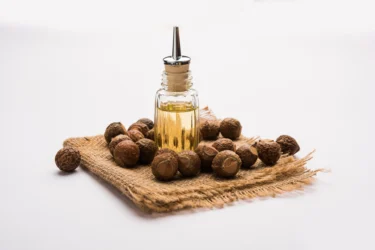
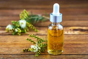
Tea tree is the name of a plant native to Australia, scientifically known as Melaleuca alternifolia1. The oil extracted from the leaves of the tea tree is tea tree oil, a widely used ingredient in a variety of household and cosmetic products, like shampoos, massage oils, laundry detergents, and skin and nail creams for its potential antiseptic benefits. Tea tree oil is pale yellow in colour. It has a strong odor and may provide a cooling sensation on the skin2. It has been widely used in Australia as a topical medicine for almost 80 years3. Common names of tea trees include tea tree oil, Australian tea tree oil, melaleuca oil and tea tree essential oil4.
The main component present in tea tree oil is terpinen-4-ol. Tea tree oil contains more than 100 compounds. However, the International Organization for Standardization has specified the main 15 components that are needed for a product to be labelled as ‘tea tree oil’. These are alpha pinene, sabinene, alpha terpinene, D-limonene, p-Cymene, 1,8-cineol, gamma terpinene, terpinolene, terpinen-4-ol, alpha terpineol, aromadendrene, ledene, delta cadinene, globulol and viridiflorol2.
Tea tree oil may have many potential uses owing to its many activities, which include
Did you know tea tree oil-based gel may aid in inflammatory gum disease called persistent gingivitis? Studies have shown that people using tea tree oil gel may have less bleeding and irritation. In my experience, a combination of tea tree oil and alpha-bisabolol, the main ingredient in chamomile, may bring down a particular strain of bacteria linked to foul breath7.
Dr. Siddharth Gupta, B.A.M.S, M.D (Ayu)
Tea trees may have potential uses for various health conditions; however, more human studies are needed to support its true scope in humans.

The overgrowth of Candida albicans in the vagina is called vaginal candidiasis or thrush; it is a fungal infection. Tea tree oil may have the potential to be developed as an effective agent for managing vaginal candidiasis. The benefits of tea tree oil in vaginal candidiasis have been observed in lab studies. However, these studies are done in laboratories and not humans; therefore, extensive clinical trials are required to check its efficacy in human beings3.

Tea tree oil may have antibacterial and anti-inflammatory properties that might be useful in managing acne. Many topical preparations containing tea tree oil have been used to stop the bacteria involved in acne. The use of tea tree oil might significantly reduce acne lesions by decreasing open and closed comedones (blackheads and whiteheads). The antiacne benefit of tea tree oil has been observed in many clinical trials, which demonstrates that topical application of tea tree oil (5%) might be effective in managing mild to moderate acne6. However, every person has a different response to different herbs. Therefore, it is better to consult a doctor before you use tea tree oil for your acne problems.

Tea tree oil was found to be beneficial in providing a cooling sensation to burn wounds and increasing the rate of wound healing6. However, this information is insufficient and requires large-scale human studies to suggest the potential uses of tea tree oil for wound healing in humans.
Tea tree oil may have potential antiviral activity against herpes simplex virus 1 and herpes simplex virus 2. Tree tea oil may have shown effectiveness in recurrent herpes labialis infection (skin rash on lips caused due to herpes virus) in a clinical trial. Tea tree oil may have also shown benefits in dealing with hand warts caused due to human papillomavirus6. However, if you have viral infections like the herpes simplex virus, it is best to seek medical help and get a proper diagnosis and treatment for your condition.

Tea tree oil is found in many preparations that are used as an alternative solution for head lice infestation. Tea tree oil, along with lavender oil, has shown effectiveness in dealing with live head lice in subjects in a clinical study. The results indicate that tea tree oil may be beneficial as an alternative method for head lice6. However, use these alternatives only under the guidance of your doctors.
Demodicisosis is an eyelid infection caused due to Demodex (a type of mite). Eyelid scrub using tea tree oil effectively eradicated eyelid demodex and helped with demodicisosis in a clinical study. Therefore, it may indicate that tea tree oil and shampoo might be beneficial in eradicating eyelid Demodex6. However, take medical help before using the tea tree for your infections and only use it if prescribed.
Though studies show the benefits of tea tree in different health conditions, these studies are insufficient and require further studies to establish the true scope of benefits of tea tree on human health. In addition, every person may respond differently to this herb. Therefore, it is essential to consult a doctor before using tea tree oil for any medical condition.
In my experience, massaging tea tree oil on the chest may relieve cough and cold symptoms, congestion, bronchitis, and other cold-related problems. Researchers believe tea tree oil has antibacterial and anti-inflammatory properties that may cope with bacterial invasion and relieve congestion and other symptoms8.
Dr. Rajeev Singh, BAMS
Your Ayurvedic physician will prescribe you the form and dose as per your requirement. However, we advise you not to replace or change your current medications with any ayurvedic or herbal preparations made from the tea tree.
The use of tea tree oil externally is generally safe. However, some people may develop skin irritation or contact dermatitis (skin inflammation) on the parts of the body where the product was used4.
However, if you see any side effects, seek immediate medical help from your physician who has prescribed it to you and get proper treatment to overcome your side effects.
Did you know the elimination of toxins like uric acid may be encouraged by tea tree essential oil? Researchers have found that tea tree essential oil may aid in pore cleaning and the removal of toxins, extra salt and water from the body through increased sweating. It may also shield your skin against the development of acne8.
Dr. Smita Barode, B.A.M.S, M.S.
Also Read: Mullein Tea: Health Benefits, Uses, Side Effects & More!
Also Read: 10 Amazing Benefits of Tea Tree Oil
There is no scientific report on the interaction between tea tree and other drugs. However, you should not assume that there are no interactions. We recommend you consult an Ayurvedic doctor; they will direct you to a better way to use it as a herb
Also Read: Turkey Berry (Solanum Torvum): Uses, Benefits and Side Effects by Dr. Rajeev Singh
Tea tree is the common name used for tea tree oil. It is the oil derived from the leaves of the tea tree plant (Melaleuca alternifolia)2,4.
Many clinical studies have shown the benefit of tea tree oil in managing acne6. However, do not use tea tree oil to self-medicate yourself; take consultations with an Ayurvedic physician.
Tea tree oil may be used as an alternative solution for head lice infestation6. However, the extent to which tea tree oil may benefit in human conditions are not known, take advice from your doctors.
Oral intake of tea tree oil should be strictly avoided, as it causes serious health issues like breathing problems, confusion, loss of muscle coordination (ataxia) and coma4.
Studies have shown that tea tree oil benefits in managing vaginal candidiasis, a vaginal infection caused by Candida albicans. But this activity has been proven in laboratory studies; further trials on humans are needed to check the efficacy of tea tree oil in human beings3. In case of any infection, it is better to consult a doctor. Self-medicating might worsen the condition.
Tea tree oil is produced by steam distillation of the leaves and terminal branches of Melaleuca alternifolia. Once the distillate is condensed, the pale-yellow oil is separated from the aqueous distillate5.
1. Hammer KA, Carson CF, Riley T v., Nielsen JB. A review of the toxicity of Melaleuca alternifolia (tea tree) oil. Food and Chemical Toxicology. 2006 May 1;44(5):616–25. Available from: https://pubmed.ncbi.nlm.nih.gov/16243420/
2. Larson D, Jacob SE. Tea tree oil. Dermatitis [Internet]. 2012 Jan [cited 2022 Mar 24];23(1):48–9. Available from: https://journals.lww.com/dermatitis/Fulltext/2012/01000/Tea_Tree_Oil.10.aspx
3. Hammer KA, Carson CF, Riley T v. In-vitro activity of essential oils, in particular Melaleuca alternifolia (tea tree) oil and tea tree oil products, against Candida spp. The Journal of antimicrobial chemotherapy [Internet]. 1998 Nov [cited 2022 Mar 24];42(5):591–5. Available from: https://pubmed.ncbi.nlm.nih.gov/9848442/
4. Tea Tree Oil | NCCIH [Internet]. [cited 2022 Mar 24]. Available from: https://www.nccih.nih.gov/health/tea-tree-oil
5. Carson CF, Hammer KA, Riley T v. Melaleuca alternifolia (Tea Tree) Oil: a Review of Antimicrobial and Other Medicinal Properties. Clinical Microbiology Reviews [Internet]. 2006 Jan [cited 2022 Mar 24];19(1):50–62. Available from: https://journals.asm.org/doi/full/10.1128/CMR.19.1.50-62.2006
6. Pazyar N, Yaghoobi R, Bagherani N, Kazerouni A. A review of applications of tea tree oil in dermatology. International Journal of Dermatology. 2013 Jul;52(7):784–90. Available from: https://pubmed.ncbi.nlm.nih.gov/22998411/
7. Pereira SL da S, Praxedes YC M, Bastos TC, Alencar PN B, da Costa FN. Clinical effect of a gel containing Lippia sidoides on plaque and gingivitis control. Eur J Dent [Internet]. 2013 Jan;7(1):28–34 [cited 2026 Jan 8]. Available from: https://pmc.ncbi.nlm.nih.gov/articles/PMC3571506/
8. Sabir S, Arshad M, Zahara K, Tabassum S, Chaudhari SK. Pharmacological attributes and nutritional benefits of tea tree oil. Int J Biosci [Internet]. 2014 Jul;5(2):80–91 [cited 2026 Jan 8]. Available from: https://www.researchgate.net/publication/279178729_Pharmacological_attributes_and_nutritional_benefits_of_tea_tree_oil_International_Journal_of_Biosciences_IJB
Disclaimer: The information provided here is for educational/awareness purposes only and is not intended to be a substitute for medical treatment by a healthcare professional and should not be relied upon to diagnose or treat any medical condition. The reader should consult a registered medical practitioner to determine the appropriateness of the information and before consuming any medication. PharmEasy does not provide any guarantee or warranty (express or implied) regarding the accuracy, adequacy, completeness, legality, reliability or usefulness of the information; and disclaims any liability arising thereof.
Links and product recommendations in the information provided here are advertisements of third-party products available on the website. PharmEasy does not make any representation on the accuracy or suitability of such products/services. Advertisements do not influence the editorial decisions or content. The information in this blog is subject to change without notice. The authors and administrators reserve the right to modify, add, or remove content without notification. It is your responsibility to review this disclaimer regularly for any changes.
2
1
Reetha, scientifically known as Sapindus mukorossi, is a large deciduous tree of the Sapindaceae family. It is commonly kReetha, scientifically known as Sapindus mukorossi, is a large deciduous tree of the Sapindaceae family. It is commonly known by many names like soapberry, soapnut, washnut, aritha, dodan, and dodani. In countries like Japan and China, Reetha has been used for centuries. In Japan, it has been used as a life-prolonging pericarp (the part of a fruit enclosing the seeds) and in China as a fruit for managing illnesses. The plant is well known for its folk traditional values1. Reetha is found in the hilly regions of the Himalayas in India. The fruit of Reetha has been used in Indian Ayurvedic medicine for decades2. Reetha is a popular ingredient of many Ayurvedic shampoos and cleansers3.
The major constituents present in Reetha are saponins, sugars, and mucilage1. The seed kernels of Reetha are a rich source of proteins and show a balanced amino acid composition as per the World Health Organization. In addition to proteins, sugars, and fibers are also present. Phytochemicals like polyphenols and saponins are also present4. The seed oil contains vitamin E and beta-sitosterol5. The nutritional value is mentioned in the table below.
Reetha may have the following beneficial properties.

Reetha contains essential compounds such as saponins. These compounds may possess anticancer and antitumour activities. Various laboratory studies have found that Reetha effectively stopped the growth of cancer cells and tumour formation in cancer cell lines. Hence, Reetha may have the potential to induce the death of cancer cells1. However, this study is insufficient because it is done in the laboratory and not on humans. Therefore, large-scale human trials are required to suggest the true potential of Reetha in fighting cancer in humans1.
According to an animal study, Reetha extract stopped the growth of the bacteria, Helicobacter pylori when administered orally. Furthermore, the extract was beneficial in clearing out the Helicobacter pylori infection in an in-vivo study1. However, this information is not enough, and further studies are required to support the potential use of Reetha in humans for managing bacterial infections.
Based on my observations, when you mix reetha with shikakai, the mixture has multiple health benefits. Together they act as antifungal, anti-bacterial, anti-inflammatory and antipyrogenic agents. Additionally, they will exhibit cleansing properties that will be highly beneficial for hair health9.
Dr. Siddharth Gupta, B.A.M.S, M.D (Ayu)
Reetha may benefit liver health. In an animal model study, the extract of the fruit pericarp of Reetha showed a positive effect on the liver of the animal. The extract might help reduce the damage caused to the liver cells1. You must consult a qualified doctor if you observe any changes in your liver health.
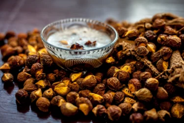
According to a lab study, the extract of Reetha might stop the growth of Candida albicans, which causes cutaneous (skin) candidiasis infection. The extract of Reetha showed strong anti-fungal activity against Candida parapsilosis. In addition, the saponin portion of the extract showed activity against the fungus Trichophyton rubrum1. This data is insufficient; therefore, extensive human studies are needed to support its benefits in humans.
According to Ayurveda, the reetha powder obtained from a reliable source might help in various skin infections like psoriasis, eczema, and pimples as it has Tridosha (Pitta, Vatta and Kapha) properties. Further, reetha decoction might have wound-healing properties and may stop secondary infections10.
Dr. Rajeev Singh, BAMS
Oil derived from the seeds of Reetha showed wound-healing benefits in animal studies. The oil also showed anti-inflammatory, antioxidant, and anti-bacterial properties. It was found that the benefit of skin wound healing may be the result of the anti-bacterial, anti-inflammatory, and antioxidant activity. The vitamin E present in the oil may provide antioxidant benefits. Reetha seed oil may have the potential for skin wound healing in humans5. However, before using Reetha for wound healing, it is better to consult a doctor to avoid further complications.

Reetha is widely used in preparations like shampoo3. The dried fruit powder may be used as a foaming agent in shampoos6. It may clean the oily secretions in the skin and might be used as a cleanser for hair and a hair tonic as it forms a natural lather1. It may also be used for removing lice from hair3. However, you should never use any herb to self-medicate yourself. It is always best to consult your doctors and only use them if recommended.
Though studies are showing the benefits of Reetha in various conditions, these are insufficient, and there is a need for further studies to establish the true extent of the benefits of Reetha on human health. Additionally, every person may respond differently to this herb. Therefore, it is essential to consult a doctor before using Reetha for any medical condition.
You should consult an Ayurvedic doctor before taking herbal supplements made of Reetha. In addition, we advise you not to replace or discontinue your ongoing medications with Ayurvedic or herbal preparations without consulting a qualified physician.
Also Read: Mehendi: Uses, Benefits, Side Effects & More!
The saponins present in the extract may be safe to be used in cosmetics7.
Therefore, if you experience any side effects, immediately rush for medical help from your doctor who has prescribed it and get proper treatment.
Also Read: Shikakai – Uses, Benefits, Side Effects & More!
There is no information on the interaction of Reetha with other drugs. However, you should not assume that there are no interactions. Therefore, it is best to follow the advice of your doctors. They will prescribe you a better way to have it as a herb.
Also Read: Castor Oil – Uses, Benefits, Precautions & More!
Reetha is a large deciduous tree, scientifically known as Sapindus mukorossi. The fruits of Reetha have been used in Ayurvedic medicines1.
The seeds of Reetha may be used as a cleanser for washing hair. It forms rich and natural lather1. Reetha may also be used to remove lice from the scalp3. However, if you have hair problems, contact a qualified doctor and ask for proper hair treatment. We recommend you do not use Reetha to self-medicate yourself.
Reetha may be used to remove the oils from the scalp generated by oily secretions1. However, it is better to seek medical help from a dermatologist if you have scalp issues.
The seeds of Reetha are edible. However, the saponins found in Reetha are bitter in taste. To remove these saponins, you can leach the seeds in running water, cook them thoroughly or change the cooking. It is not advisable to consume large quantities of Reetha as it can be toxic8. Therefore, it is recommended to take the advice of an Ayurvedic doctor; they will guide you with the best form and dosage to have Reetha as a herb.
The common names of Reetha aresoapnut, soapberry, aritha, washnut, dodan, and dodani1.
Powdered seeds may be used in the management of dental caries1. It is not advisable to self-medicate and consult a dentist if you face any dental problems. Self-medicating might worsen the situation.
Reetha seed oil may have the potential for skin wound healing as it showed wound healing benefits in animal studies. The beneficial property of skin wound healing may be due its anti-bacterial, anti-inflammatory and antioxidant activity5. In case of any skin wound, consult a doctor. Self-medicating might worsen the situation.
1. Upadhyay A, Singh DK. Pharmacological effects of Sapindus mukorossi. Revista do Instituto de Medicina Tropical de Sao Paulo [Internet]. 2012 Sep [cited 2022 Mar 17];54(5):273–80. Available from: https://pubmed.ncbi.nlm.nih.gov/22983291/
2. Chaudhary SK, Mandal AB, Bhar R, Gopi M, Kannan A, Jadhav SE, et al. Effect of graded levels of soapnut (Sapindus mukorossi) shell powder on reproductive performance in broiler breeders. Asian-Australasian Journal of Animal Sciences [Internet]. 2019 [cited 2022 Mar 17];32(1):118. Available from: https://www.ncbi.nlm.nih.gov/pmc/articles/PMC6325405/
3. (PDF) Sapindus mukorossi (areetha): An overview [Internet]. [cited 2022 Mar 17]. Available from: https://www.researchgate.net/publication/267698560_Sapindus_mukorossi_areetha_An_overview
4. Chavan RS, Rathod VK. Evaluation of nutritional and medicinal potential of defatted Sapindus mukorossi seed kernel. Preparative biochemistry & biotechnology [Internet]. 2022 [cited 2022 Mar 21];52(1):56–61. Available from: https://pubmed.ncbi.nlm.nih.gov/33881946/
5. Chen CC, Nien CJ, Chen LG, Huang KY, Chang WJ, Huang HM. Effects of Sapindus mukorossi Seed Oil on Skin Wound Healing: In Vivo and in Vitro Testing. International Journal of Molecular Sciences [Internet]. 2019 May 2 [cited 2022 Mar 21];20(10). Available from: https://www.ncbi.nlm.nih.gov/pmc/articles/PMC6567820/
6. Lodha G. Formulation and Evaluation of Polyherbal Shampoo to Promote Hair Growth and Provide Antidandruff Action. Journal of Drug Delivery and Therapeutics [Internet]. 2019 Aug 30 [cited 2022 Mar 21];9(4-A):296–300. Available from: https://www.researchgate.net/publication/335843667_Formulation_and_Evaluation_of_Polyherbal_Shampoo_to_Promote_Hair_Growth_and_Provide_Antidandruff_Action
7. Du M, Huang S, Zhang J, Wang J, Hu L, Jiang J. Toxicolological Test of Saponins from Sapindus mukorossi Gaerth. Open Journal of Forestry [Internet]. 2015 [cited 2022 Mar 21];05(07):749–53. Available from: https://www.scirp.org/journal/paperinformation.aspx?paperid=60418
8. Sapindus mukorossi Chinese Soapberry, Soap Berry, Chinese Soapberry, Soapnut Tree PFAF Plant Database [Internet]. [cited 2022 Mar 21]. Available from: https://pfaf.org/user/Plant.aspx?LatinName=Sapindus+mukorossi
9. Patil SB, Pawar VV. The Review Article of Medicinal Uses Reetha and Shikakai. Int J Multidiscip Res. 2022;4(6):1-4. Available from: https://www.ijfmr.com/papers/2022/6/983.pdf
10. Mishra P, Maury S. The review on herbal hair mask for the prevention of dandruff. World J Pharm Res. 2022;11(6):343-351. doi:10.20959/wjpr20226-24107. Available from: https://wjpr.s3.ap-south-1.amazonaws.com/article_issue/6be932c05223675930031c2aab378899.pdf
Disclaimer: The information provided here is for educational/awareness purposes only and is not intended to be a substitute for medical treatment by a healthcare professional and should not be relied upon to diagnose or treat any medical condition. The reader should consult a registered medical practitioner to determine the appropriateness of the information and before consuming any medication. PharmEasy does not provide any guarantee or warranty (express or implied) regarding the accuracy, adequacy, completeness, legality, reliability or usefulness of the information; and disclaims any liability arising thereof.
Links and product recommendations in the information provided here are advertisements of third-party products available on the website. PharmEasy does not make any representation on the accuracy or suitability of such products/services. Advertisements do not influence the editorial decisions or content. The information in this blog is subject to change without notice. The authors and administrators reserve the right to modify, add, or remove content without notification. It is your responsibility to review this disclaimer regularly for any changes.
7
1
Trigonella foenum-graecum or Methi (fenugreek) is one of the oldest traditional medicinal plants belonging to the Fabaceae family. It is cultivated in the Indian subcontinent, North Africa, and the Mediterranean region. Methi is used in many forms, such as herb (dried leaves), fresh leaves (vegetables), and spice (seeds). Fenugreek is nutritious, functional, and nutraceutical in nature. All these traits make fenugreek a well-known spice crop used in human diets[1].
Fenugreek is a rich source of dietary fibres and other nutrients needed for proper growth. Fenugreek is also rich in several phytochemicals such as alkaloids, carbohydrates, amino acids, minerals and steroidal saponins[2].
Nutritional value of Methi (fenugreek):
In addition, Methi also contains minerals such as potassium, calcium, magnesium, zinc, copper, iron, and manganese. Beta carotene and Vitamin C are considered essential components of Methi.
Methi possesses many properties, as mentioned below.
Because of a wide variety of active phytochemicals, methi may show potential uses against many disease conditions. Some of the potential uses of methi are mentioned below.

Rheumatoid Arthritis is a disease that affects the joints by causing chronic inflammation, characterised by tissue injury and synovial proliferation. As per animal studies[4], supplementing fenugreek mucilage could inhibit oedema by lowering the activity of inflammatory enzymes. Properties of fenugreek may help in managing arthritis symptoms. However, you must strictly adhere to the doctor’s advice and treatment when dealing with arthritis. Reach out to your doctor before using any herbal remedy.

A compound called protodioscin in methi could show anticancer properties against cancer cell lines. Fenugreek extract also showed inhibition of the growth of breast and pancreatic cancer cell lines in studies. As per animal studies[6], the administration of fenugreek extracts also helps reduce tumour cell growth. However, more research is required to support the use of fenugreek against cancer in humans. Therefore, fenugreek should not be used against cancer without consulting a healthcare provider first.
The gel fraction of fenugreek may help avoid gastric lesions. The stomach protective benefit of fenugreek might be due to the presence of flavonoids and polysaccharides in the gel portion of fenugreek. In addition, the anti-secretory activity of fenugreek might play a protective role against ulcers. However, if you are struggling with stomach problems like ulcers, talk to your doctor and get a proper diagnosis and treatment[5].

The components present in methi might be effective against neurological disorders like Parkinson’s disease, Alzheimer’s disease, and depression. For example, fenugreek extract may help lessen depression as it inhibits the activity of the monoamine oxidase enzyme. In addition, components like flavonoids present in fenugreek might help reduce behavioural abnormalities by positively altering the pathways and expressions of proteins and enzymes. However, any brain disorders should be met with proper diagnosis and treatment. If you are having trouble with any brain functions, talk to your healthcare provider and receive help[7].

Asthma is a lung disorder characterised by inflamed airways that restrict the pathway for inhalation and exhalation. According to a study[8], fenugreek extract was effective in treating mild asthma and can be recommended as adjuvant therapy. Daily use of methi may help avoid the symptoms of asthma. However, do not use methi as an alternative to asthma treatment. You should talk to a doctor before making any decisions.

Methi may help manage weight without any significant side effects. Many animal studies have shown the effectiveness of methi in weight loss. Methi’s high amount of fibre might help suppress hunger. Consumption of methi powder was found to be helpful in reducing weight, as per animal studies[9]. The galactomannan present in methi seeds may help in excreting the sugars from the body before they are absorbed into the blood. However, before making any changes to your diet, you are advised to talk to your healthcare provider or dietitian.
Extract of the fenugreek seeds may provide protective benefits against alcohol-induced liver damage. The polyphenolic compounds present in the extract may show liver-protective benefits. The fenugreek seeds might help protect the liver cells and function from the toxic effects of alcohol. However, taking herbal remedies if you are suffering from any liver condition may worsen the situation. Therefore, you need to consult your healthcare provider before using any herbal remedy for the liver[10].
Though studies show the benefits of methi in various conditions, these are insufficient, and there is a need for further studies to establish the true extent of the benefits of methi on human health.
There are many ways to make use of methi in daily life.
You must consult a doctor before taking methi or any herbal supplements. Do not discontinue or replace an ongoing treatment of modern medicine with an Ayurvedic/herbal preparation without consulting a doctor.
The side effects associated with methi use are given below.
Before taking methi or any other herbal remedy, you need to talk to your doctor about the possible side effects of the herbal treatment. This will help you avoid unwanted side effects.
Also Read: Sesame Seeds: Benefits, Uses, Side Effects and More!
Here are some general precautions you need to take while using methi.
Also, you should talk to a doctor, who will help you make well-informed choices.
Also Read: What to Eat and What to Avoid in Appendicitis?
Methi may interact with some medicines and alter their activity.
If you are taking medicines for any health conditions, talk to your doctor about the possible interactions of the treatment with other herbs and medicines. It will help you avoid unwanted herb-medicine interactions.
Also Read: 15 Amazing Health Benefits of Turmeric Milk!
The consumption of Methi has been associated with weight loss. In addition, the high fibre content of Methi may suppress hunger, thereby promoting weight loss. Make sure to talk to your doctor or dietician before making any changes to your diet or taking any herbal remedy for weight loss.
The paste of Methi leaves can be applied to the scalp before the bath. It helps in hair growth, preserves natural colour, helps reduce dandruff, and helps keep hair silky.
Methi has shown effectiveness in managing diabetes, as per several animal studies[12]. It could increase insulin sensitivity and improve the condition of insulin resistance. However, human trials are awaited to confirm its efficacy in human beings. Therefore, it should not be used as an alternative or replacement for conventional diabetes therapy.
Using fenugreek in pregnant women should be avoided as it may lead to abortion. It is also associated with birth defects in the offspring. So, the use of fenugreek should be strictly avoided during pregnancy. It is better to talk to a doctor before using it during pregnancy.
Methi seeds can be used as a spice. Methi seeds are well-known for their aromatic, spicy properties and are commonly used as a seasoning. However, talk to your doctor before using methi as a herbal remedy.
Disclaimer: The information provided here is for educational/awareness purposes only and is not intended to be a substitute for medical treatment by a healthcare professional and should not be relied upon to diagnose or treat any medical condition. The reader should consult a registered medical practitioner to determine the appropriateness of the information and before consuming any medication. PharmEasy does not provide any guarantee or warranty (express or implied) regarding the accuracy, adequacy, completeness, legality, reliability or usefulness of the information; and disclaims any liability arising thereof.
66
7